Impactful Findings from Medical Imaging Research

Dive into a collection of short summaries of recent medical imaging studies that shed light on hot topics such as structured reporting, texture analysis, radiomics, or novel imaging biomarkers. Identify the role mint Lesion™ played in these studies and contact us if you have any questions about our software platform.

Here you will find a detailed list of scientific publications in which mint Lesion™ has had a significant impact:
Explore the publications

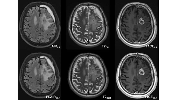
University Hospital Tübingen: Neuro-Oncological Imaging with Deep Learning Reconstruction (DLR)

A recent study conducted by the University Hospital of Tübingen investigated the potential of deep learning reconstruction (DLR) in magnetic resonance…

Screenshot of structured reading template for NSCLC Staging

Software-Assisted Structured Reporting Improves TNM Classification Accuracy in NSCLC Staging

In this multi-center collaboration, thoracic radiology experts developed and evaluated a software-assisted structured reporting (SR) framework for…

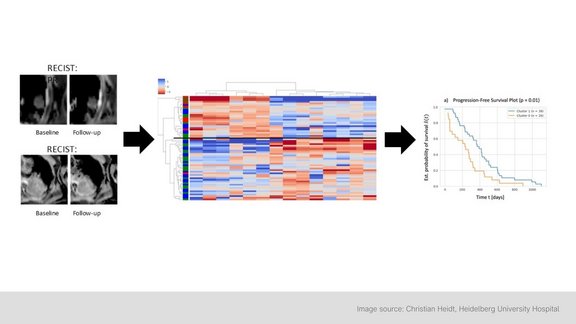
Image shows graphic related to study's research on iRECIST and RECIST

University Hospital Cologne: Comparison of iRECIST and RECIST 1.1 for Evaluating Immunotherapy in Melanoma and Non-Small Cell Lung Cancer

A retrospective study conducted at University Hospital Cologne compared two criteria for assessing therapeutic response to immunotherapy: iRECIST and…

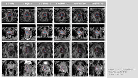
Image showing MR image analysis using the dedicated semiautomatic software tool mint Lesion™

LMU Klinikum Munich: Monitoring Prostate Cancer Treatments with VTP and HIFU - The Use of Multiparametric MRI

The prospective study conducted by LMU Klinikum Munich investigates the treatment of localized prostate cancer using two techniques: vascular-targeted…

The image serves as a graphical abstract, displaying visuals from the study, ranging from MRI scans to the analysis of delta-radiomics texture features

Heidelberg University Hospital: Delta-Radiomics Features from ADC Maps as Early Predictors of Treatment Success in Lung Cancer Therapy

In this prospective study conducted by Heidelberg University Hospital, researchers investigated whether changes in radiomic features from…

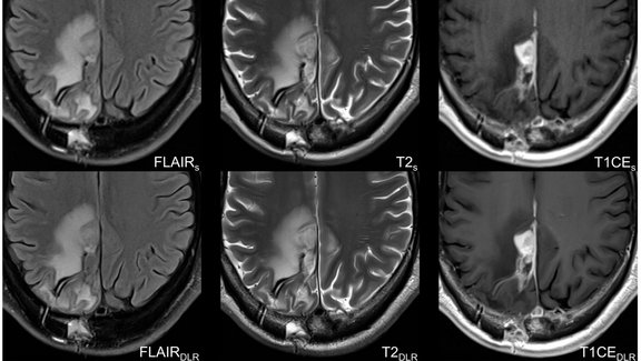
Three important sequences (FLAIR, T2, T1 with contrast agent) in the assessment of glioblastoma

University Hospital Tübingen: Advancing MRI Efficiency in Glioblastoma Care with Deep Learning

This study explores the use of deep learning (DL) to optimize MRI protocols for glioblastoma patients. Glioblastomas, known for being the most…

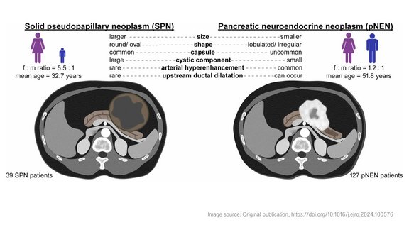
Illustrations of solid pseudopapillary neoplasms and pancreatic neuroendocrine neoplasms

Heidelberg University Hospital: Key Imaging Features to Distinguish Solid Pseudopapillary Neoplasms from Pancreatic Neuroendocrine Neoplasms

Solid pseudopapillary neoplasms (SPNs), or Frantz tumors, are rare pancreatic tumors accounting for 2-3% of all pancreatic neoplasms. These tumors…

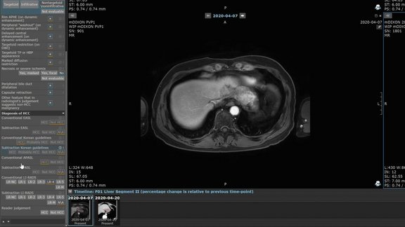
mint Lesion screenshot with HCC diagnosis according to APASL, AASLD, LI-RADS, KLCA-NCC, and EASL guidelines

Multicentric Study: Comparison of Diagnostic Guidelines for Hepatocellular Carcinoma

Recent advancements in MRI techniques and tumor biology have led to updated hepatocellular carcinoma (HCC) diagnostic guidelines from various liver…

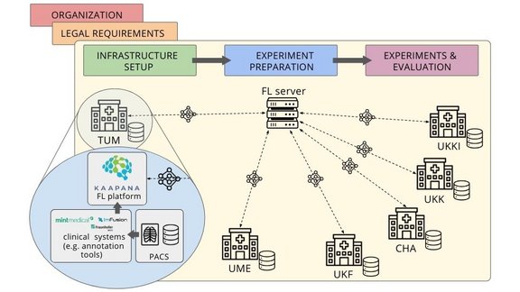
Schematic visualization of the federated learning study and its data infrastructure

RACOON: A Guide to Bridging the Gap Between Simulated and Real-World Federated Learning Research

Deep learning (DL) has become an important part of radiological image analysis. To train these deep-learning models, access to large and diverse…

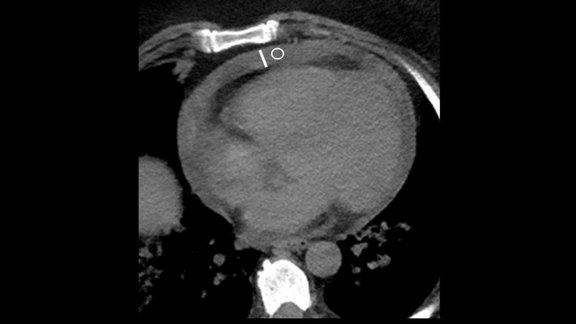
Picture shows a scan of pericardial effusion

RACOON: Study emphasizes the clinical relevance of pericardial effusion as an imaging biomarker in COVID-19 patients

A multicenter study [1] based on the German research infrastructure project RACOON (Radiological Cooperative Network of the COVID-19 pandemic; a Netzw…

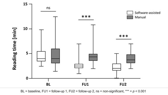
Diagram that shows reduced reading times for the mint Lesion™ approach at both follow-ups.

UKE Hamburg: Study Shows that Software-Assisted Assessments Enhance iRECIST Evaluation

This research study [1] aimed to compare the feasibility and reliability of manual versus software-assisted assessments of computed tomography (CT)…

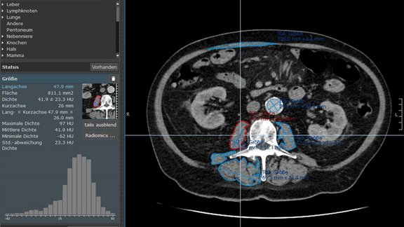
mint Lesion screenshot with segmented muscles and radiomics

University Hospital Ulm: CT Radiomics and Machine Learning in Sarcopenia Evaluation for Esophageal and Gastric Cancer Patients

A recent study [1] conducted by researchers at University Hospital Ulm explored the role of sarcopenia in patients with esophageal or gastric cancer.…