Deep learning (DL) has become an important part of radiological image analysis. To train these deep-learning models, access to large and diverse datasets is required. While the use of a centralized data lake would be most convenient, this approach is often complicated by privacy regulations.
Federated Learning (FL) facilitates collaborative model training without centralizing data. Despite its potential, however, most FL studies remain in simulated environments due to practical challenges, with limited real-world implementations. As a result, real-world FL projects (especially in radiology) are rare. Additionally, these few existing initiatives often neglect to communicate their insights on overcoming these hurdles, thus leading to a significant knowledge gap.
This study [1] wants to bridge the gap between simulated and real-world FL research. It reviews FL literature and details the development of an FL infrastructure within the Netzwerk Universitätsmedizin (NUM) project German Radiological Cooperative Network (RACOON). The RACOON project is dedicated to leveraging artificial intelligence (AI) to enhance research in radiological diagnosis and therapy across 38 German university hospitals. As a part of this initiative, each hospital is equipped with the structured radiological reporting software, mint Lesion™.
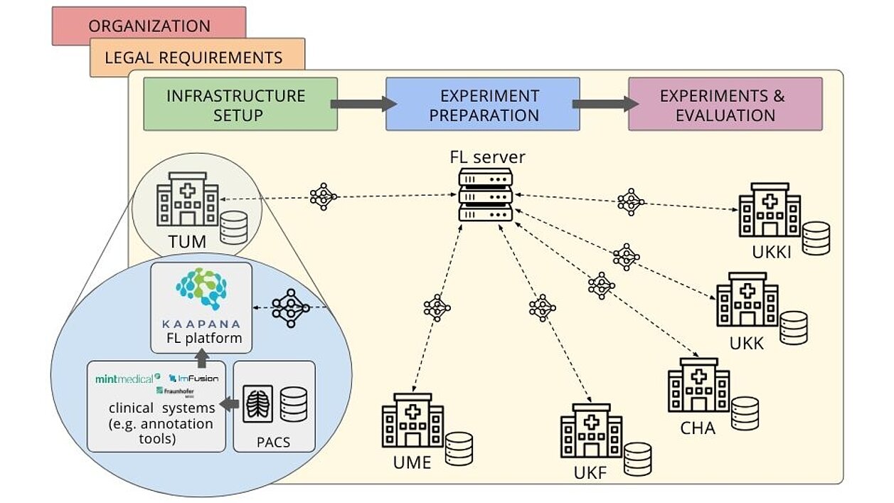
Regarding their study, the researchers state: “Within the framework of the German Radiological Cooperative Network (RACOON), we have built and established the first nation-wide FL initiative of its kind, that includes all 38 university hospitals of the country. We evaluated its functionality by conducting real-world FL experiments to collaboratively train DL segmentation models on radiological image data across six university hospitals. This setup provides a ready-to-use infrastructure for future researchers to conduct clinical research using FL without having to build their own systems. During developing this FL initiative and conducting experiments with real-world datasets in a real-world setting, we encountered and overcame numerous difficulties and practical hurdles.”
The FL system was tested by training FL models on lung pathology segmentation across six university hospitals. For this purpose, radiological images were queried from the PACS and then processed and annotated by, inter alia, mint Lesion™, to be then trained using the FL platform. Furthermore, FL's performance was compared to simpler alternatives, like local model training and ensembling approaches.
Results show that FL outperforms these less complex alternatives, highlighting its potential value in real-world applications. The study also offers a comprehensive guide for establishing FL initiatives, emphasizing the importance of strategic organization, robust data management, and infrastructure. It aims to help future researchers implement FL in clinical settings, demonstrating its superiority and addressing real-world implementation challenges.
Read the original publication here.
[1] Bujotzek, Markus R., Ünal Akünal, Stefan Denner, et al., Real-World Federated Learning in Radiology: Hurdles to Overcome and Benefits to Gain, Preprint, 2024.