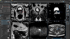
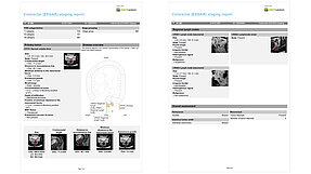
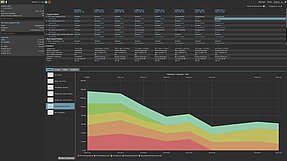
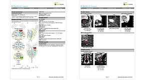
From Images to Data and

with Data to Knowledge

Our innovative solution seamlessly integrates context knowledge with image information, catering to both structured reporting in clinical routine and the specific requirements of clinical trials. The easy-to-use radiology software provides assisted data capture, rendering all image information - incl. radiomics or tumor growth rate - readily accessible

Key benefits at a glance

Lung
Collect, structure and
link the imaging data,
assessments, meta-data and
the images themselves
Pancreas
Assure consistency,
compliance, and accessibility
of both imaging and clinical
data
Pancreas
Improve interdisciplinary
communication by connecting
data from multiple sources

Use Scenarios

mint Lesion™ is a radiology platform designed for a variety of applications, including routine clinical practice, clinical trials, and quantitative biomarker imaging research. It can be effortlessly configured to meet specific requirements in any context 

Our live demos are just as flexible as mint Lesion™ itself.
We will customize your demo to your interests and a time that suits you.

loading...

What our users say about mint Lesion™

mint Lesion™ allows to efficiently conduct precise oncological treatment monitoring and communicate comprehensible reports to the clinical colleagues with a simple key stroke.

PD Dr. Felix NensaUniversity Hospital Essen

mint Lesion™ is an asset for the professionality and motivation of radiological tasks. Since the introduction of this software we have structured our evaluation procedure for clinical trials and are experiencing an effective improvement in the daily routine of oncological therapy evaluation.

Prof. Dr. Ulf TeichgräberUniversity Hospital Jena

mint Lesion™ is an innovative tool which helps radiologists in becoming central in oncologic multidisciplinary teams.

Prof. Dr. Andrea LaghiSapienza University of Rome

mint Lesion™ is an outstanding software that facilitates cancer proforma reporting and streamlines tumor treatment response assessment. Its functionality and versatility help radiologists do a better job!

Prof. Dow-Mu KohThe Royal Marsden NHS Foundation Trust

Accurate, reproducible, and standardized assessment of treatment response is of fundamental importance in oncology. A powerful yet easy to use and integrated software such as mint Lesion™ may be exactly what it takes to migrate such a capability from the world of clinical trials to everyday practice of Radiology and Oncology.

Prof. Dr. Dominik FleischmannStanford University

mint Lesion™ allows for fast and adequate TNM staging and provides an excellent schematic overview of an individual patient’s disease for oncologists, surgeons, and the patients.

PD Dr. Thorsten PersigehlUniversity Hospital Cologne

mint Lesion™ knows about the partially complex subtleties of assessment criteria, which makes the assessment of lesions a lot easier compared to conventional workstations.

Prof. Dr. Philippe PereiraSLK Clinics Heilbronn

Good integration in the existing IT was important to us – appropriate interfaces to RIS and PACS were implemented quickly and professionally.

Prof. Dr. Elmar KotterUniversity Hospital Freiburg

By using mint Lesion™, we are able to generate standardized, complete and consistent oncologic reports. Not only can we describe all the important aspects of a particular tumor, but we are also storing important data in the background, which can be used for quality control and science.

Dr. Remy GeenenNoordwest Ziekenhuisgroep Alkmaar

The mint Lesion™ COVID-19 template is a tool that helps me and my colleagues to collect all important information about the patients – both clinical and imaging data. It is detailed and ensures a comprehensive standardized report.

Dr. Konstantinos StathopoulosInstitut Jules Bordet Bruxelles

mint Lesion™ has become an indispensable part of our tumor boards and clinical trials. It significantly strengthens the recognition of radiology and the cooperation between imaging departments and with clinical partners.

Prof. Meinrad BeerUniversity Hospital Ulm

The Mint Medical team has worked tirelessly with Imperial College London to develop and support an entire bespoke software platform to host the NIHR funded MROC study, from patient registration to the end of study.  Co-ordinating CT and multiparametric MRI images, blinded double reads, treatment plans, surgical and histopathology findings and outcomes on the MROC platform has transformed the way we can deliver image-based clinical trials and bio-banking of study data.

Prof. Andrea RockallImperial College London

Events

BRG 2023
10/05 to 10/07/2023 LANDSHUT, GERMANY

Annual Congress of the Bavarian Society of Radiology

SABI Annual Meeting 2023
10/07 to 10/11/2023 DALLAS, TX, USA

Innovation towards professional, financial, and environmental sustainability.

RANZCR Brisbane 2023
10/19 to 10/21/2023 BRISBANE, AUSTRALIA

73rd Annual Scientific Meeting

News

Early lung cancer detection through LDCT screening as a potential way to improve patient outcomes in Germany

The establishment of a nation-wide lung cancer screening program in Germany has been a subject of considerable interest and debate among healthcare…

RACOON FHIR Workshop: Empowering Healthcare Research via Enhanced Interoperability

This week Brainlab hosted the RACOON FHIR Workshop with around 40 participants. The workshop was organized by Mint Medical and supported by Snke OS,…

Tumor Growth Rate Modeling: A Novel Approach to Evaluating the Efficacy of Cancer Therapies

The 2020 review of Clinical Trial Evidence Supporting US Food and Drug Administrative Approval of Novel Cancer Therapies Between 2000 and 2016…