Recent advancements in MRI techniques and tumor biology have led to updated hepatocellular carcinoma (HCC) diagnostic guidelines from various liver study associations. Conducted across 11 South Korean hospitals, this study aims to compare the diagnostic performance of four guidelines and readers’ judgement in diagnosing HCC using an automatic algorithm in high-risk patients.
The study was conducted retrospectively and involved 2,237 patients at risk for HCC with 2,445 focal liver lesions (FLLs). Approximately 69.3% of the FLLs were HCC, and 78.4% of the patients had chronic hepatitis B. Among the FLLs, 31.3% were smaller than 20 mm, with 59.1% of these being HCC.
Focal liver lesions were reviewed according to four guidelines: American Association for the Study of Liver Diseases (AASLD)/Liver Imaging Reporting and Data System (LI-RADS), Korean Liver Cancer Association– National Cancer Center (KLCA-NCC), European Association for the Study of the Liver (EASL), and Asian Pacific Association for the Study of the Liver (APASL). The guidelines were assessed based on their accuracy, sensitivity, specificity, positive predictive value, and negative predictive value.
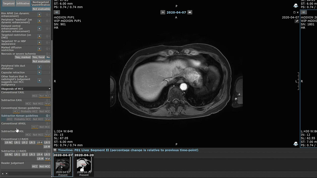
Two of the authors collaborated with the Mint Medical development team to create a mint Lesion™ template incorporating common imaging features and precise automatic diagnostic assignments from the four guidelines (AASLD/LI-RADS, KLCA-NCC, EASL, and APASL).
“This collaboration aimed to implement a seamless automatic diagnostic process before finalizing the electronic case report form,” stated the authors. “A total of 46 questionnaires were provided for each FLL, addressing major features, targetoid and nontargetoid LR-M features, the presence of tumor in vein (LR-TIV category), and ancillary features.”
The comparison of diagnostic performance in all focal lesions showed that guidelines of the Asian Pacific Association for the Study of the Liver (APASL) had the highest sensitivity at 89.1%, followed by those of the Korean Liver Cancer Association–National Cancer Center (KLCA-NCC) at 78.2%, of the American Association for the Study of Liver Diseases (AASLD)/Liver Imaging Reporting and Data System (LI-RADS) at 70.5%, and of the European Association for the Study of the Liver (EASL) at 69.3%.
AASLD/LI-RADS exhibited the highest specificity at 89.6%, followed by EASL at 88.1%, KLCA-NCC at 84.1%, and APASL at 52.2%.
For small FFLs, APASL had higher sensitivity (85.0%), whereas AASLD/LI-RADS had the highest specificity (91.6%). The accuracy for diagnosing HCC in small lesions was higher using Eastern guidelines (APASL and KLCA-NCC) compared to Western guidelines (AASLD/LI-RADS and EASL).
Diagnostic performance was also analyzed in subgroups with cirrhosis and with chronic hepatitis B without cirrhosis. The subgroup analysis revealed that the performance of the diagnostic guidelines varied based on specific patient characteristics and tumor sizes. The Western guidelines displayed high specificity, while the Eastern guidelines demonstrated high sensitivity across both subgroups.
Interobserver agreement for key imaging features used in HCC diagnosis varied, with some features showing moderate to substantial agreement among radiologists.
“[U]sing four hepatocellular carcinoma diagnosis guidelines, we confirmed that Eastern guidelines exhibited high sensitivity and Western guidelines demonstrated high specificity,” concluded the researcher. “The Korean Liver Cancer Association–National Cancer Center guidelines achieved the highest accuracy and the American Association for the Study of Liver Diseases/Liver Imaging Reporting and Data System guidelines displayed the highest diagnostic odds ratio.“
The study highlights the strengths and limitations of each guideline, emphasizing the importance of tailored diagnostic approaches and the potential for human expertise to enhance diagnostic accuracy.
Read the original publication here.
Yoon, JH, Kim YK, Kim JW et al. Comparison of Four Diagnostic Guidelines for Hepatocellular Carcinoma Using Gadoxetic Acid–enhanced Liver MRI, Radiology 2024.