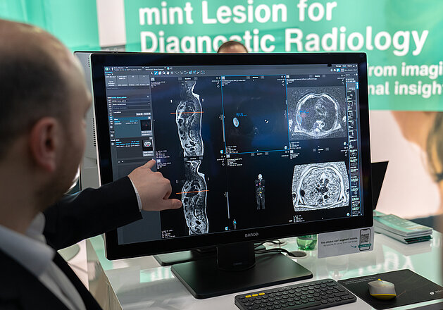
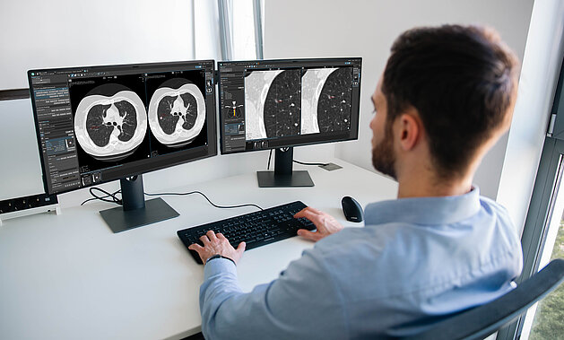
RadMag Special Edition: Onur Özek on the Challenges of Lung Cancer Screening in Germany
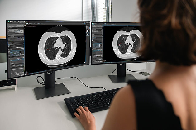
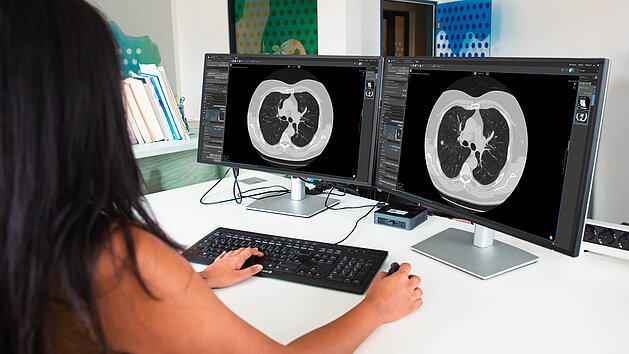
With the nationwide rollout of Germany’s lung cancer screening, radiology providers are facing new challenges. Structured reporting, mandatory second…
Read more