jump to content jump to footer

Streamline Prostate Cancer Screenings with Prostate.Carcinoma.ai in mint Lesion

The Prostate.Carcinoma.ai plug-in, developed by our partner FUSE-AI, is a powerful addition to mint Lesion, designed specifically to enhance prostate cancer screenings. This advanced solution leverages artificial intelligence to automate and accelerate prostate MR evaluations based on the PI-RADS 2.1 guidelines, transforming the workflow and precision for radiologists.

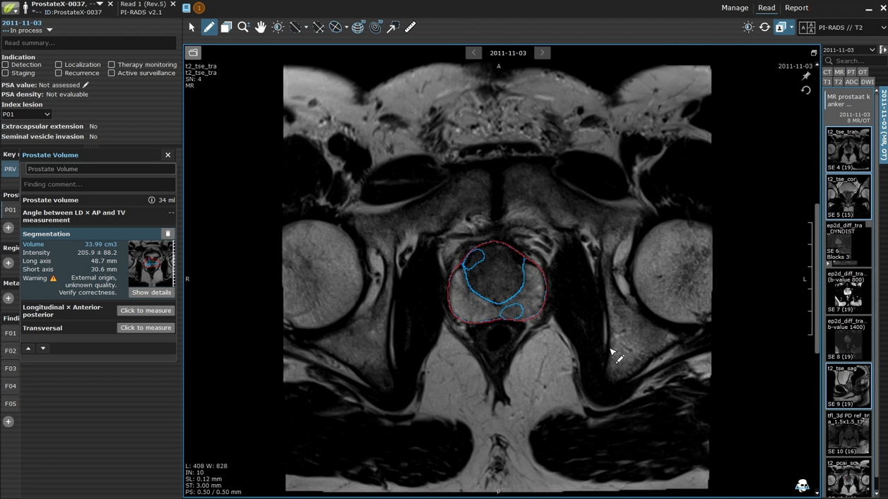
At the click of a button, Prostate.Carcinoma.ai performs full segmentation of the prostate and its anatomical zones, and detects and segments potentially malignant lesions. mint Lesion maps each finding onto the standardized prostate lesion scheme, eliminating the need for time-consuming manual lesion annotation and drawing.

Radiologists maintain full control over the AI-generated results, with the ability to review, reject, modify, or add new findings to ensure accuracy. Based on these inputs, mint Lesion automatically calculates the PI-RADS score, delivering a comprehensive and structured report.

mint Lesion also enables longitudinal tracking of prostate lesions for MRI-based active surveillance. Radiologists can easily monitor changes in lesions over time, supporting informed decision-making for patient management and enhancing the continuity of care.

Learn more on our partner page.

From PCWG3 to PCWG4: Evolving Standards in Prostate Cancer Clinical Trials

The Prostate Cancer Working Group 4 (PCWG4) updates and extends the recommendations of PCWG3 to reflect a patient-centric approach, the emergence of…

Read more
Mint Medical and mediaire strengthen the prostate cancer care pathway with AI-Powered MRI Solutions

Mint Medical, a Snke company, and mediaire announce a collaboration to advance AI‑powered MRI workflows through the interoperability of mint Lesion…

Read more
Medical professional reviewing patient's MRI images on computer monitor with patient in MRI machine in background.
TGSE-BLADE demonstrated significantly reduced geometric distortion and artifacts caused by intracranial air, enabling more accurate detection of ischemic changes and improving diagnostic confidence in early postoperative MRI.
Postoperative Brain MRI After Tumor Resection: Reducing Artifacts and Improving Diagnostic Accuracy

Postoperative MRI after brain tumor surgery is often affected by artifacts caused by intracranial air, limiting reliable image interpretation.

A…

Read more
scroll-top