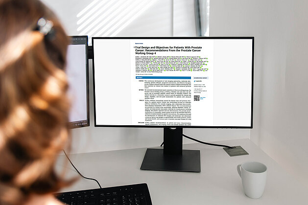
Tumor response assessment in neuro-oncology clinical trials requires careful attention to measurement protocols and confirmation scan requirements. To address evolving research needs and standardize assessment across different tumor types, the Response Assessment in Neuro-Oncology Criteria (RANO) 2.0 criteria proposes several significant differences compared to the previous standards, including RANO for high-grade gliomas (RANO-HGG), low-grade gliomas (RANO-LGG), modified RANO (mRANO), and immunotherapy RANO (iRANO).
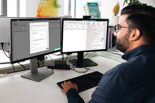
mint Lesion now supports RANO 2.0 criteria, offering researchers a structured approach to implementing these guidelines. The platform provides both default RANO 2.0 settings and the flexibility to adapt parameters.
Configurable Assessment Parameters
mint Lesion provides default settings based on RANO 2.0 guidelines, with options to adjust parameters according to your study requirements.
Available Configuration Options
Adapt mint Lesion to match your study protocol:
- Measurement Approach: Select two-dimensional measurements, volumetric analysis, or both
- Tumor Type Definition: Specify contrast-enhancing tumors, non-enhancing tumors, or mixed tumors for your study, or allow the definition of tumor type per case
- Confirmation Scan Protocol: Apply the default 12-week confirmation window for enhancing tumors post-radiotherapy, modify the timeframe, or disable/enforce confirmation scans for all timepoints
- Response Categories: Configure major and minor response classifications for non-enhancing tumors
- Threshold Parameters: Use RANO 2.0 standard thresholds or apply custom values
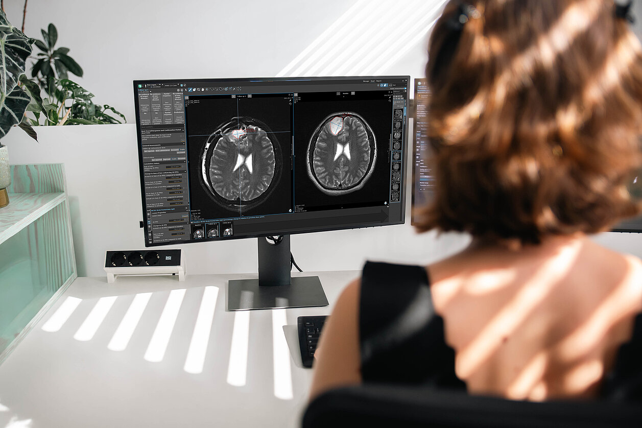
Tumor Burden Calculation and Tracking
mint Lesion calculates tumor burden using:
- Sum of Products of Perpendicular Diameters (SPPD) for two-dimensional measurements
- Sum of Volumes (SOV) for volumetric analysis
The software tracks changes relative to multiple reference points:
- Baseline
- Previous timepoint
- Nadir (lowest tumor burden)
- Nadir after pseudoprogression (lowest point at or after the first preliminary progression when confirmation scans are required)
Automated Confirmation Scan Logic
mint Lesion applies confirmation scan requirements according to RANO 2.0 defaults or custom study settings. The software determines when confirmation is needed and identifies potential pseudoprogression cases. The visualization feature allows tracking of tumor burden changes over time.
Detailed Measurement Breakdown
mint Lesion displays calculated tumor burden values organized by lesion type and measurement method:
- SPPD for enhancing lesions
- SPPD for non-enhancing lesions
- SOV for enhancing lesions
- SOV for non-enhancing lesions
The calculations account for target lesions, non-target lesions, and new lesions according to RANO 2.0 criteria. Target Lesion Response is determined based on cumulative values and shown separately for enhancing/non-enhancing lesions and 2D/3D measurements. Appropriate new lesions and progressive non-target lesions are automatically added to the tumor burden.
RANO 2.0 Reference Publications
Implementation in mint Lesion takes into account these publications:
- Wen PY, van den Bent M, Youssef G, et al. RANO 2.0: Update to the Response Assessment in Neuro-Oncology Criteria for High- and Low-Grade Gliomas in Adults. J Clin Oncol. 2023;41(33):5187-5199. doi:10.1200/JCO.23.01059
- Ellingson BM, Sanvito F, Cloughesy TF, et al. A Neuroradiologist's Guide to Operationalizing the Response Assessment in Neuro-Oncology (RANO) Criteria Version 2.0 for Gliomas in Adults. AJNR Am J Neuroradiol. 2024;45(12):1846-1856. Published 2024 Dec 9. doi:10.3174/ajnr.A8396
- Sakata A, Fushimi Y, Oshima S, et al. RANO 2.0: critical updates and practical considerations for radiological assessment in neuro-oncology. Jpn J Radiol. 2025;43(10):1557-1574. doi:10.1007/s11604-025-01821-6
Interested in seeing how mint Lesion handles RANO 2.0 implementation for your study?
Schedule a live demonstration to explore the configuration options, automated calculations and reporting features. Our team can walk you through specific scenarios relevant to your research needs.