Stay Informed: Transforming Radiology with Structured Reporting and Data-Driven Approaches

Dive into our activities, projects, and product updates. Catch on the latest industry news and learn who we are as a company and as a team.

Doctors looking at MRI scans to evaluate a glioblastoma.

Optimizing Glioblastoma Imaging: Enhancing MRI Efficiency and Quality with Deep Learning

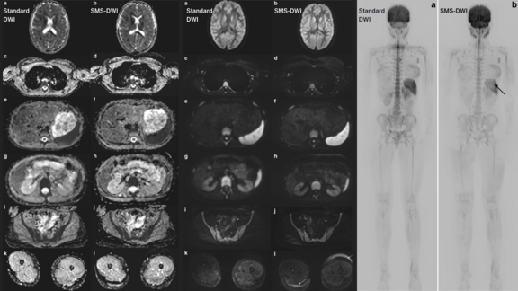
This study investigates the use of deep learning (DL) to optimize MRI protocols for glioblastoma patients, aiming to reduce scan time and improve image quality.

Glioblastomas are aggressive brain tumors requiring frequent MRI monitoring, which can be challenging due to lengthy scan times and motion artifacts. Traditional methods to shorten scan times, like parallel acquisition techniques (PAT) and compressed sensing (CS), have limitations such as reduced signal-to-noise ratio and overly smooth images.

The study, involving 33 patients, found that DL-optimized MRI sequences reduced scan time by 30% while enhancing image quality and maintaining diagnostic accuracy. These improvements are particularly beneficial for patients who struggle with lengthy MRI procedures, offering a promising advancement in glioblastoma care.

Read more about the study here.

Related Resources

Related Resources

Potential of using structured data from clinical trials and routine for AI and radiomics research

Dr. Nils Grosse Hokamp shares his experience of using mint Lesion™ at the University Hospital Cologne in this brief interview. He talks about how they…

Mint Medical provides a reading template for the standardized assessment and documentation of COVID-19 disease (coronavirus) based on CT imaging

In the first week of March, we will make the relevant software functions available for all our current users at no cost. We plan to offer the COVID-19…

Clear, consistent, and complete documentation of imaging-derived information

The radiology report is expected to provide information that impacts life changing treatment decisions at every cancer care step in which imaging is…