Structured data in radiology is crucial for accurate diagnoses and treatment planning and serves as the basis for detailed clinical practice and research analysis.
We spoke to Prof. Dr. Thorsten Persigehl, Professor of Oncological Imaging at the Faculty of Medicine and Head of Oncological and Abdominal Imaging at the Institute of Diagnostic and Interventional Radiology at the University Hospital of Cologne, about the challenges and opportunities of setting up a comprehensive oncological imaging database in the radCIO project.
Prof. Persigehl, can you give us a brief overview of the radCIO?
radCIO stands for the Radiology Center for Integrated Oncology, within the Center for Integrated Oncology (CIO) at Cologne University Hospital. We founded the radCIO out of the radiology department to respond to the specialization in radiology with ever-improving imaging possibilities, increasing knowledge of molecular genetics and new therapy options in oncology. Our primary goal is to enhance the care of our cancer patients with ultimately better outcomes. Therefore, we aim to optimize radiological processes and digitalization to provide comprehensive, structured imaging data for patient care and clinical research.
Currently, imaging data in clinics is often stored in an unstructured way, which means that it cannot be used directly for comprehensive clinical or experimental research purposes. It was, therefore, crucial for us to have a modern IT platform that efficiently archives structured data in a digital database while integrating it into the clinical context and enabling direct digital exchange to the other oncology systems, such as via HL7 and FHIR. We have built a digital network with a robust data structure and an extensive "real-world" database accessible for further analysis and future research questions. It is essential for evidence-based medicine that we incorporate this information directly into future research questions and make it available to oncologists for their investigations.
Which role does mint Lesion™ play in the radCIO and how does the software platform contribute to data-based radiology?
mint Lesion™ ultimately forms the backbone for the entire radCIO.
In general, the radCIO is based on an EU tender, REACT-EU, and several challenges arose when looking for a suitable platform solution:
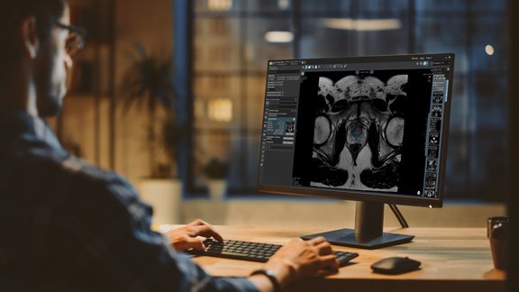
We were looking for an IT platform that would enable fully digitized storage of radiology data, particularly image-based and structured. The annotations should be available directly in the image to make the data accessible not only for additional quantitative analyses, such as radiomics, but also for future research questions and artificial intelligence (AI) applications. There should be the possibility of AI integration.
We wanted a digital platform with a dashboard to analyze the structured data in real-time. At the same time, interfaces, particularly a FHIR interface, should be integrated to enable us to collaborate seamlessly with internal and external clinical and research partners, including as part of tumor boards and with the cancer registry. Cancer patients and their oncologists should have personal access to their evaluations in a patient portal. In addition, the data should also be available directly in pseudo- and anonymized form for research collaborations.
We were able to implement all these functions with our Mint platform, which enables us not only to answer current questions in oncological imaging but also to address future research questions and be ready for clinical applications of AI solutions.


