A prospective, monocentric study [1] conducted by Vanessa F. Schmidt and her colleagues at the University Hospital Munich (LMU) evaluated the morphological and volumetric changes in MRI data of patients with symptomatic benign prostate syndrome (BPS) following prostatic artery embolization (PAE).
Benign prostatic hyperplasia (BPH) is the most common prostate condition in men over the age of 50 and patients suffer not only from complications, but also from a significant impairment of quality of life. A variety of minimally invasive procedures have been developed to alleviate the symptoms of benign prostatic hyperplasia when medications are ineffective. Among these, PAE has been established as a low-risk procedure that offers high technical success rates and improvement in qualitative and quantitative measurements of lower urinary tract symptoms.
“Regarding changes in prostate MRI over time after PAE, only manual assessments have been performed to date, both with respect to volumetric characteristics and morphologic changes in SIs [signal intensities],” declared the authors. “Thus, we aimed to investigate the semi-automated approach as an alternative method to assess prostate MRI after PAE, including the identification of potential benefits and the correlation with clinical parameters.”
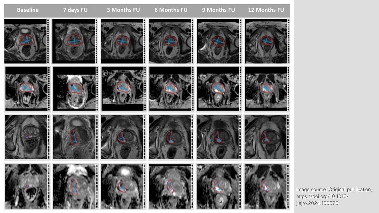
MRI and clinical data sets from 27 patients with symptomatic BPS were assessed before and at one, six, and 12 months after PAE. mint Lesion™ was used:
- to semi-automatically segment both the central (CG) and the total gland (TG),
- to assess the changes in signal intensities on T1-weighted (T1w), T2-weighted (T2w), and apparent diffusion coefficient (ADC)-map and
- to analyze volumetric changes (TGV, CGV) and intra-vesical prostatic protrusion (IPP). In addition to the semi-automatic volumetry of TGV, the ellipse formula described by Sosna et al. [2] was used to calculate the TGV volumetry.
TGV significantly decreased at each timepoint after PAE as assessed by both semi-automatic approach and ellipse formula (p = 0.025; p <= 0.05). CGV was significantly reduced one month after PAE (p = 0.038), but there were no significant differences in CGV between baseline and six or 12 months after PAE.
All patients with demonstrated IPP at baseline (92.6%) experienced a significant decrease of IPP volume at each timepoint (p = 0.028; p = 0.010; p = 0.018).
A significant decrease in ADC values of the CG was observed one month after PAE (p < 0.001), while there were no significant differences in Tw1 and Tw2 signal intensities between baseline and any time point.
“The semi-automatic approach is an adequate method for assessing volumetric and morphological changes in prostate MRI following PAE because significant differences in ADC values can be measured and there is no need for manual identification of infarct areas,” concluded the researchers.
[1] Schmidt, V.F., Schirren, M., Heimer, M.M., Kazmierczak, P.M., Cyran, C.C., Wildgruber, M., Seidensticker, M., Ricke, J., Solyanik, O. Semi-Automatic MRI Feature Assessment in Small- and Medium-Volume Benign Prostatic Hyperplasia after Prostatic Artery Embolization. Diagnostics 2022. https://doi.org/10.1016/j.ejrad.2020.109242
[2] Sosna, J., Rofsky, N.M., Gaston, S.M., DeWolf, W.C., Lenkinski, R.E. Determinations of prostate volume at 3-Tesla using an external phased array coil: Comparison to pathologic specimens. Acad Radiol. 2003