Stay Informed: Transforming Radiology with Structured Reporting and Data-Driven Approaches

Dive into our activities, projects, and product updates. Catch on the latest industry news and learn who we are as a company and as a team.

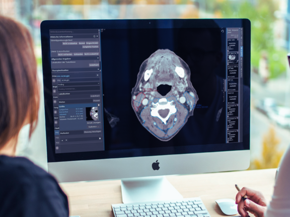
A picture of radiologists looking at a screen showing the restaging of the HNSCC

Study Highlights the Potential of Radiomics in Medical Intervention

A recent study conducted by the Medical University of Innsbruck highlights the potential of radiomics in exploring the impacts of Radiochemotherapy (RCT) on patients with Head and Neck Squamous Cell Carcinoma (HNSCC). This study is the first to explore short-term RCT-induced skeletal muscle alterations after RCT in HNSCC patients applying radiomics, a data-driven approach aiming at extracting and processing quantitative data to analyze image-based information.

With the help of the mint Lesion™ Radiomics module, the research examined radiation-induced changes in the sternocleidomastoideus muscle (SCM) and paravertebral musculature (PVM) post-RCT. Analyzing key radiomic features like volume, mean positivity of pixels, and uniformity the study team was able to confirm the optimal window of 6 to 12 weeks for salvage surgery post-RCT, minimizing the risk of complications due to tissue fibrosis. These findings further showcase how radiomics can enhance medical treatment strategies and indicate a bright path for ongoing research in this area.
Read more about the study

Related Resources

Related Resources

The image serves as a graphical abstract, displaying visuals from the study, ranging from MRI scans to the analysis of delta-radiomics texture features

Heidelberg University Hospital: Delta-Radiomics Features from ADC Maps as Early Predictors of Treatment Success in Lung Cancer Therapy

In this prospective study conducted by Heidelberg University Hospital, researchers investigated whether changes in radiomic features from…

Doctors looking at MRI scans to evaluate a glioblastoma.

Optimizing Glioblastoma Imaging: Enhancing MRI Efficiency and Quality with Deep Learning

This study investigates the use of deep learning (DL) to optimize MRI protocols for glioblastoma patients, aiming to reduce scan time and improve…

Three important sequences (FLAIR, T2, T1 with contrast agent) in the assessment of glioblastoma

University Hospital Tübingen: Advancing MRI Efficiency in Glioblastoma Care with Deep Learning

This study explores the use of deep learning (DL) to optimize MRI protocols for glioblastoma patients. Glioblastomas, known for being the most…