
Research Insights from Dr. Madelaine Hettler: The Template Designer in Use
We spoke with Dr. Madelaine Hettler, resident physician at the Sarcoma Center of University Medical Center Mannheim. Together with Prof. Dr. med. Jens Jakob, she coordinates the RACOON-SAGA project within the Network University Medicine (NUM). The project aims to improve the pre-therapeutic characterization of soft tissue sarcomas and thereby provide a stronger foundation for optimized therapy decisions.
How and in what context have you used the Template Designer?
Within the RACOON-SAGA project in the Network University Medicine (NUM), we have been working extensively with the Mint Medical Template Designer. Our project focuses on evaluating MRI data together with clinical and prognostic information, and for this purpose, the Template Designer provides an excellent and well-structured platform.
Specifically, we use the Template Designer to design our project on soft tissue sarcomas. The goal was to analyze functional parameters from MRI images while also systematically collecting clinical data. The Template Designer allows us to combine all of this information within a clear structure: from measurements in imaging to parameters such as patient age, tumor subtype, and therapies applied.
How do you rate the user-friendliness of the Template Designer?
Overall, I found it very pleasant to work with the Template Designer. Of course, it takes a little time at the beginning to get familiar with the functions, but afterward, it proved to be very intuitive and user-friendly. You don’t necessarily have to be a radiologist to use it—I was able to work with it very comfortably.
What I found particularly helpful was that all the relevant measurements for imaging were already available and could simply be inserted. For the clinical data, we had multiple options as well, including both multiple-choice and free-text questions. This made it possible to capture everything we needed for our project. The multiple-choice options, in particular, made data entry easy and efficient: users just have to click through, which speeds up and simplifies the process considerably.
What overall impression did you gain while working with the Template Designer?
The Template Designer offers a clear and well-organized platform, providing an overview of both imaging and clinical data at any time—which I found very valuable. In my view, this is its main advantage: the ability to collect clinical data in parallel with imaging data and directly relate the two.
The measurements were very straightforward to handle. Even as a surgeon, I found it easy to navigate and could perform the measurements just as I had envisioned. One specific measurement was not available at first, but together with the Mint team, we quickly found a solution. Now, all the measurements we need are available, allowing us to use the Template Designer to its full potential.

Our conversation with Dr. Hettler demonstrates how the Template Designer can bridge the gap between imaging and clinical data in research projects, making an important contribution to structured data collection.
We sincerely thank Dr. Hettler for sharing her insights and experiences with the Template Designer.
If you’d like to learn more about the possibilities of the Template Designer or explore how structured data could support your research or clinical workflows, feel free to reach out to us.
Dr. med. Hettler is a resident physician at the Department of Surgery at University Medical Center Mannheim. Together with Prof. Dr. med. Jens Jakob, she coordinates the RACOON-SAGA project. For her scientific work, she was awarded the 2024 Research Prize of the German Sarcoma Foundation. For questions about the RACOON-SAGA project, you can contact the team at sarkomchirurgie@umm.de.
Related Resources
Related Resources
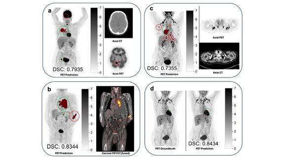
LMU University Hospital: Artificial Intelligence for TNM Staging in NSCLC – How Reliable Are AI-Based Segmentations?
The recent study “Artificial intelligence for TNM staging in NSCLC – a critical appraisal of segmentation utility in [¹⁸F]FDG PET/CT” provides a…
Successful “RECIST and Beyond” Workshop in Cologne: Advancing Precision in Oncologic Imaging
How can complex tumor findings be assessed accurately, reproducibly, and in line with clinical guidelines?
RACOON – Imaging, Data & Collaboration for Better Decisions
Modern radiology faces a central question: how can imaging and clinical data be combined in a way that leads to more precise diagnoses,…