Free-text report often lacks both content and clarity, burdening referring physicians with the time-consuming and cumbersome task of finding and extracting essential information. The crucial details may either be buried in paragraph statements or left out due to requisite concision in reporting. These communication barriers often cause conflicts between the perceived and the intended meaning of the report and endanger the precision of diagnosis.
According to a recent study published in JAMA Otolaryngology - Head & Neck Surgery [1], there is a discordance between clinician-perceived and radiologist-intended meaning of the postradiotherapy PET/CT freeform report for Head and Neck Cancer. The study results suggest that oncologists' perception of patient response from such freeform reports is unreliable and doesn't consistently reflect the radiologist's intended meaning, which is strongly associated with patient survival. According to the study results, discord existed in around one-third of the considered cases. The study thereby concluded that the use of a standardized reporting system may improve communication between clinicians and radiologists and increase the value of HNSCC post-RT PET/CT reports.
Such interdisciplinary misunderstandings are disadvantageous and potentially harmful for the patients as they may cause failure to diagnose and adequately treat progressive disease or lead to unnecessary follow-up procedures.
Although accurate communication by radiologists of patient response is essential, there is no universally accepted standardized method for such communication. Therefore there is a strong need to improve the quality of radiology reporting, which is one of the problems we at Mint Medical seek to solve with mint Lesion™.
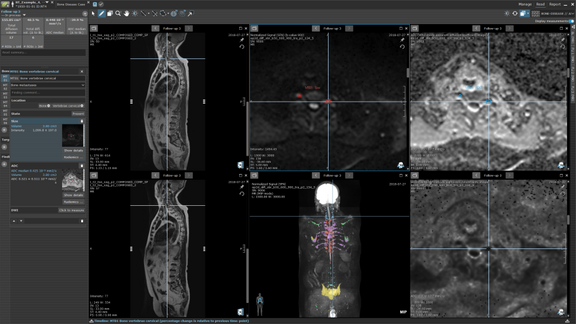
Whether in a clinical routine or a clinical trial setting, mint Lesion™ reports are standardized, criteria-compliant, and comprehensive. With mint Lesion™, radiologists can transform images into a digital stream of highly structured and quantitative diagnostic data. Structured reports generated by mint Lesion™ substantially alleviate referring physicians' cognitive burden, saving time and supplying them with reliable information that fosters data-driven diagnosis and therapy decisions.
Request a sample copy of a mint Lesion™ structured report here https://mint-medical.com/contact
[1] Patel Z, Schroeder JA, Bunch PM, et al. Discordance Between Oncology Clinician–Perceived and Radiologist-Intended Meaning of the Postradiotherapy Positron Emission Tomography/Computed Tomography Freeform Report for Head and Neck Cancer. JAMA Otolaryngol Head Neck Surg. Published online August 18, 2022. doi:10.1001/jamaoto.2022.2290