Stay Informed: Transforming Radiology with Structured Reporting and Data-Driven Approaches

Dive into our activities, projects, and product updates. Catch on the latest industry news and learn who we are as a company and as a team.

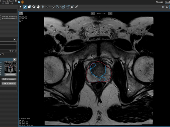
Screenshot of a prostate lesion in mint Lesion

Streamline Prostate Cancer Screenings with Prostate.Carcinoma.ai in mint Lesion

The Prostate.Carcinoma.ai plug-in, developed by our partner FUSE-AI, is a powerful addition to mint Lesion, designed specifically to enhance prostate cancer screenings. This advanced solution leverages artificial intelligence to automate and accelerate prostate MR evaluations based on the PI-RADS 2.1 guidelines, transforming the workflow and precision for radiologists.

At the click of a button, Prostate.Carcinoma.ai performs full segmentation of the prostate and its anatomical zones, and detects and segments potentially malignant lesions. mint Lesion maps each finding onto the standardized prostate lesion scheme, eliminating the need for time-consuming manual lesion annotation and drawing.

Radiologists maintain full control over the AI-generated results, with the ability to review, reject, modify, or add new findings to ensure accuracy. Based on these inputs, mint Lesion automatically calculates the PI-RADS score, delivering a comprehensive and structured report.

mint Lesion also enables longitudinal tracking of prostate lesions for MRI-based active surveillance. Radiologists can easily monitor changes in lesions over time, supporting informed decision-making for patient management and enhancing the continuity of care.

Learn more on our partner page.
Related Resources

Related Resources

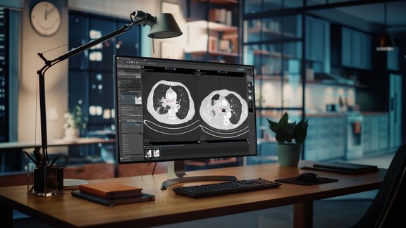
Virtual examination of human lungs on a modern interface screen

Early Detection of Treatment Response in Lung Cancer Using Delta-Radiomics Features

The study conducted by Heidelberg University Hospital investigates the use of diffusion-weighted MRI (DWI) to predict early treatment outcomes in…

The image serves as a graphical abstract, displaying visuals from the study, ranging from MRI scans to the analysis of delta-radiomics texture features

Heidelberg University Hospital: Delta-Radiomics Features from ADC Maps as Early Predictors of Treatment Success in Lung Cancer Therapy

In this prospective study conducted by Heidelberg University Hospital, researchers investigated whether changes in radiomic features from…

Doctors looking at MRI scans to evaluate a glioblastoma.

Optimizing Glioblastoma Imaging: Enhancing MRI Efficiency and Quality with Deep Learning

This study investigates the use of deep learning (DL) to optimize MRI protocols for glioblastoma patients, aiming to reduce scan time and improve…