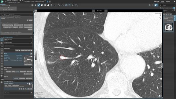
The benefits of structured oncologic reporting with mint Lesion™

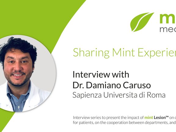
Dr. Damiano Caruso of Sapienza Universita di Roma recounts how he got to know mint Lesion™ at a workshop hosted by the ESOI. Among other workstations, mint Lesion™ automatically stood out by being easy to use, practical, and “very consistent between the different timepoints of oncological examination.”
 
He describes how at their oncologic center, mint Lesion™ is used for clinical trials and for clinical routine, as the software provides them with standardized, structured reports which contain all necessary data needed for complex oncological cases.

Click here or on the image above to watch the full video on YouTube.
Related Resources

Related Resources

Doctors looking at MRI scans to evaluate a glioblastoma.

Optimizing Glioblastoma Imaging: Enhancing MRI Efficiency and Quality with Deep Learning

This study investigates the use of deep learning (DL) to optimize MRI protocols for glioblastoma patients, aiming to reduce scan time and improve…

Three important sequences (FLAIR, T2, T1 with contrast agent) in the assessment of glioblastoma

University Hospital Tübingen: Advancing MRI Efficiency in Glioblastoma Care with Deep Learning

This study explores the use of deep learning (DL) to optimize MRI protocols for glioblastoma patients. Glioblastomas, known for being the most…

Picture of Dr. Maurice Heimer, radiology resident

Insights into the BORN Project: Development and Successes with Dr. Maurice Heimer

The BORN-project of the Bavarian Center for Cancer Research (BZKF) is making swift progress in its second funding phase. A key aspect of the project…