How to improve reading procedures and reduce errors in clinical trials with imaging endpoints

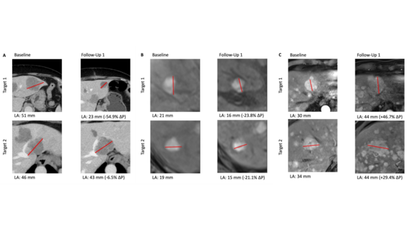
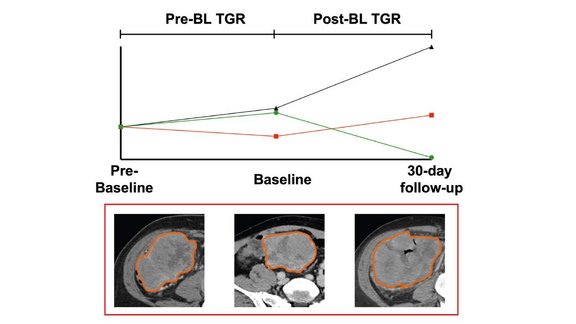
Dr. Laura Oleaga from Hospital Clinìc de Barcelona explains how she and her team have benefitted from implementing mint Lesion™ in the face of an increasing volume of clinical trials. She highlights how standardized reading procedures and reports limit the possibility of errors in clinical trial work, improve the communication between radiology and oncology, and deliver the necessary documentation for trial audits.

 
Moreover, she decsribes the potential that arises by using mint Lesion™ in their clinical routine – extending clinical trial excellence to the routine care of each oncological patient.

Related Resources

Related Resources

RECIST measurements of a patient with uveal melanoma and hepatic metastases

University Hospital Tuebingen: Study compares the Efficacy of SIRT and CS-PHP in Uveal Melanoma with Hepatic Metastasis

A study[1] conducted by researchers at University Hospital Tuebingen retrospectively compared the efficacy of two liver-targeted therapies,…

Study Analyzing the Tumor Growth Rate as a Prognostic Biomarker in Lymphoma Patients Under CAR T-Cell Therapy

University Hospital Munich (LMU): Study Analyzing the Tumor Growth Rate as a Prognostic Biomarker in Lymphoma Patients Under CAR T-Cell Therapy

A recent study[1] conducted by researchers at the University Hospital Munich (LMU) investigated the role of tumor growth rate (TGR) in predicting the…

A picture of a person looking at a brain scan and the interface of mint Lesion™  on a computer

Advancing Neuro-Oncology Assessments: Mint Medical to Integrate Newly Published RANO 2.0 Criteria into mint Lesion™

Glioblastomas and other gliomas are the most common malignant primary brain tumor types, yet few effective therapies are available. Clinical research…