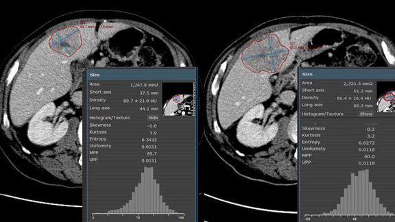
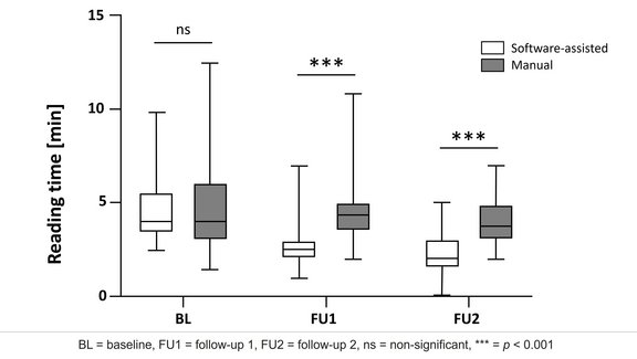
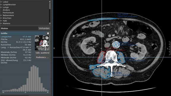
In a retrospective study [1], a team from Royal Marsden in London and Sutton explored changes of CT texture analysis metrics in unresectable liver metastases in patients with colorectal cancer (CRC) treated with Cetuximab monotherapy.
“Targeted therapy is nowadays widely used in many oncological settings, such as metastatic colon cancer, even as first-line therapy. Due to its mechanism, the effectiveness of this drug regimen is not necessarily related to a shrinkage of neoplastic lesions, therefore we are in need of new radiological biomarkers that can better assess and possibly predict patient outcome. Texture analysis is an emerging tool that can extrapolate quantitative data reflecting the grey-level distribution of the lesion’s voxels. Our aim is to take one step further towards better understanding which textural metrics could be used in assessing response to targeted therapy,” Dr. Edoardo Raimondi explained.
Baseline and first-assessment contrast enhanced CT images of 30 CRC patients were retrospectively reviewed. The researchers compared results of responders (Complete Response or Partial Response) and non-responders (Stable Disease or Progressive Disease) defined at best-response according to RECIST 1.1 throughout treatment. Within the patient cohort, 16 patients were responders and 14 non-responders. Four patients in each group had a single liver metastasis, all other patients had at least two.
mint Lesion™ was used to extract and record area- and volume-based first-order texture parameters of up to two hepatic metastases, as well as to automatically assess their percentage change from baseline to first assessment.
At baseline, no differences in texture parameters were observed between responders and non-responders. At first assessment, the average area metrics in responders showed lower entropy (6.06 vs 6.36; p=0.006) and higher uniformity (0.02 vs 0.014; p=0.002) compared to non-responders. Responders showed a percentage change of -2.08% in area-entropy and of -1.89% volume-entropy, significantly different (p < 0.05) from non-responders who showed +1.09% change in area-entropy and +0.91% change in volume-entropy. The percentage changes in area-uniformity and volume-uniformity in responders was +8.10% and +8.09% compared to -4.51% and -3.92% in non-responders (p>0.05).
“Given the differences observed in area and volume-entropy and uniformity between responders and non-responders, these metrics could be used as predictive imaging biomarker of response,” the authors concluded. Further studies should confirm these data and identify a threshold for early prediction of response.
[1] Early prediction of response of CT textural analysis in unresectable liver metastases of colorectal cancer treated with Cetuximab chemotherapy
E. Raimondi, S. Picchia, K. Kouvelakis, K. Khan, D. Cunningham, D.-M. Koh, M. A. Bali


