Stay Informed: Transforming Radiology with Structured Reporting and Data-Driven Approaches

Dive into our activities, projects, and product updates. Catch on the latest industry news and learn who we are as a company and as a team.

New iRECIST Oncologic Response Criteria already in clinical use at the University Medical Center – Tübingen, Germany

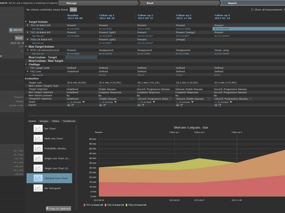
Shortly following the release of the new iRECIST- oncologic response criteria (Lancet Oncology, March 2017) the University Medical Center in Tübingen, Germany had already begun to implement this new response criteria into their clinical routine using mint LesionTM. The iRECIST guidelines are intended to provide a standard approach to the evaluation of solid tumors with measurements and assessment of the disease burden in trials where an immunotherapy is used.

PD Dr. Bernhard Klumpp who provides radiological response evaluation of of studies is one of the first radiologists to apply the new iRECIST response assessment in mint LesionTM. “The possibility of transferring the existing report process to an iRECIST process is of course sensational. For this a new measurement is not necessary, one simply changes perspective during observation of the process. Moreover, special features of iRECIST are indicated in the application”, says Dr. Klumpp.

The new iRECIST response evaluation platform is now available for all mint LesionTM Users. iRECIST is expected to be widely applied in both routine radiology evaluation as well as for application as a standardized response assessment methodology as required in oncology clinical trials.
Related Resources

Related Resources

Puzzle pieces connecting to an interoperable system

Healthcare on FHIR: Igniting the Potential of Interoperability

Interoperability plays a crucial role in healthcare: it enables seamless communication of patient information across different systems, leads to…

Doctors looking at MRI scans to evaluate a glioblastoma.

Optimizing Glioblastoma Imaging: Enhancing MRI Efficiency and Quality with Deep Learning

This study investigates the use of deep learning (DL) to optimize MRI protocols for glioblastoma patients, aiming to reduce scan time and improve…

Three important sequences (FLAIR, T2, T1 with contrast agent) in the assessment of glioblastoma

University Hospital Tübingen: Advancing MRI Efficiency in Glioblastoma Care with Deep Learning

This study explores the use of deep learning (DL) to optimize MRI protocols for glioblastoma patients. Glioblastomas, known for being the most…