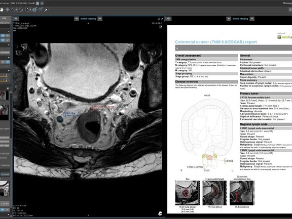
The radiology report is expected to provide information that impacts life changing treatment decisions at every cancer care step in which imaging is required. However, recent literature demonstrates that very often this information is disregarded.
The current clinical practice shows that reports consistently omits information that is associated with clinical outcome and risk of local and distant tumor recurrence. “Crucial information needed to make therapeutic decisions were missing from 52% of radiology cancer reports” evaluated in a multicenter study [1].
Some alarming examples: Studies analyzing rectal cancer reports show that the extramural venous invasion (EMVI) was provided in only 7.7 % [2] and the tumor involvement of the mesorectal fascia (MRF) was missed in one third [3] of free-text reports.
Another study [4] shows that the TNM stage was mentioned explicitly in only 6% of the text-based reports and in 22%, information was incomplete.
mint Lesion guides users through the reading process and ensures that all relevant information is collected in accordance with e.g. ACR-RADS classification, TNM guidelines or criteria for therapy assessment. It enables a clear and consistent documentation of the imaging information and significantly improves the completeness of the findings.
1 Patel A, Rockall A, Guthrie A, et al Can the completeness of radiological cancer staging reports be improved using proforma reporting? A prospective multicentre non-blinded interventional study across 21 centres in the UK, BMJ Open 2018;8:e018499.
2 Sahni, V. A., Silveira, P. C., Sainani, N. I. and Khorasani, R. Impact of a Structured Report Template on the Quality of MRI Reports for Rectal Cancer Staging, American Journal of Roentgenology. 2015;205: 584-588.
3 Brown, P.J., Rossington, H., Taylor, J. et al. Standardised reports with a template format are superior to free text reports: the case for rectal cancer reporting in clinical practice. Eur Radiol 29, 5121–5128 (2019).
4 Sexauer, R., Weikert, T., Mader, K. et al. Towards More Structure: Comparing TNM Staging Completeness and Processing Time of Text-Based Reports versus Fully Segmented and Annotated PET/CT Data of Non-Small-Cell Lung Cancer. Contrast Media & Molecular Imaging (2018) Article ID 5693058.


