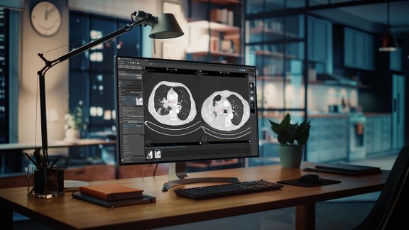
The Clinical Research Imaging Core (CRIC) of the University of Wisconsin Carbone Cancer Center (UWCCC) recently published a poster at the AACI CRI conference titled “Shared Investment to Build a Strong, Streamlined, and Accessible RECIST Foundation in Clinical Research”. On this poster, core manager Mr. Alex Arbuckle and faculty lead Dr. Steve Cho discuss that the previous tumor response assessment service they were using was “inflexible, cumbersome, did not align with research aspirations, and was cost prohibitive.”.
Through a joint review of various solutions, they arrived at the decision to implement mint Lesion as their centralized tumor response assessment system. The joint investment and implementation of mint Lesion by the UWCCC and UW Department of Radiology Medical Imaging Research Support has greatly improved the inclusion of imaging expertise in the assessments. Since then, it has “improved cost effectiveness, continually improved assessment time to completion, and has allowed for quick start-up and navigation of investigator initiated trials.” In the first 9 months, they were able to expand from 5 users in 2 programs to 12 users in 6 programs and halved the time required for completing an assessment.
Read the full poster by clicking here: Link to AACI CRI poster


