Emily Ferris and Alex Arbuckle describe how using mint Lesion™ has enhanced their clinical trial operations, enabling “a more cooperative approach between [their] radiologists and oncologists” while simultaneously saving time and reducing costs.
Faced with a multitude of clinical trial site reads each day, Ms. Ferris and Mr. Arbuckle explain how they benefit from a more efficient workflow by using a dedicated software platform which provides a comprehensive and compliant response assessment and supports them in maintaining oversight of clinical trial imaging analysis design.
Ms. Ferris is a Lead Image Analyst and Mr. Arbuckle is a Clinical Research Imaging Core Manager at the University of Wisconsin Carbone Cancer Center.
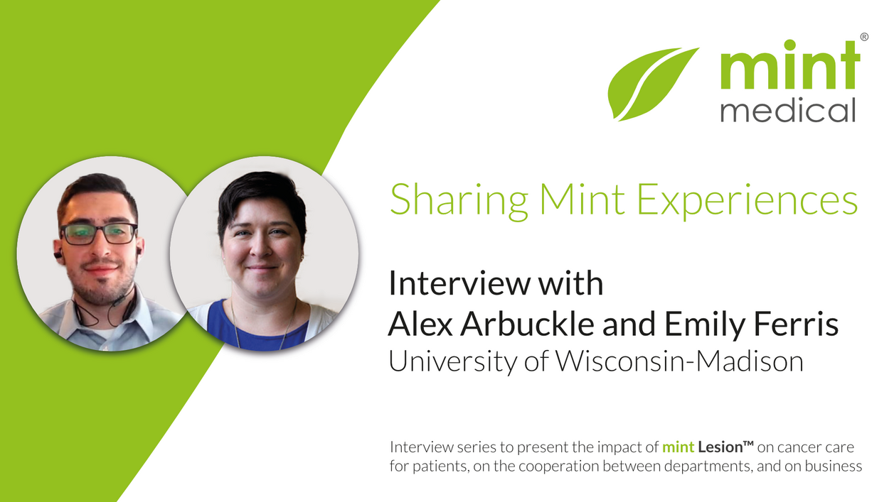
Enhancing flexibility and communication while saving costs and time in clinical trials
Related Resources
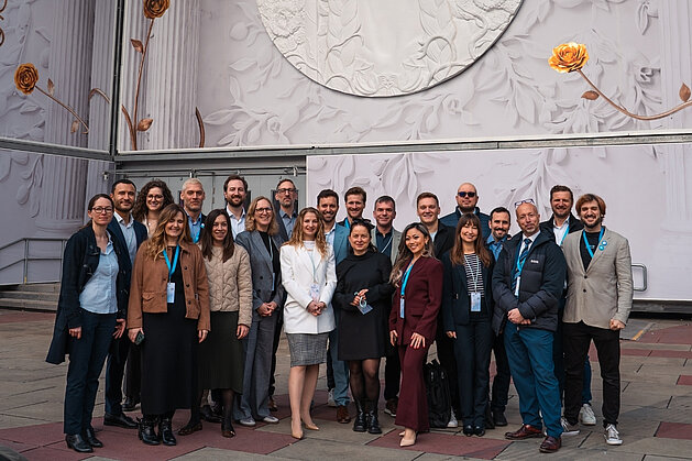
Mint Medical and mediaire strengthen the prostate cancer care pathway with AI-Powered MRI Solutions
Mint Medical, a Snke company, and mediaire announce a collaboration to advance AI‑powered MRI workflows through the interoperability of mint Lesion…
Read more
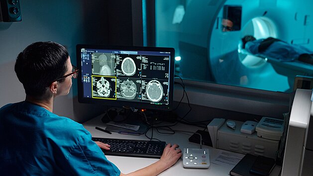
Postoperative Brain MRI After Tumor Resection: Reducing Artifacts and Improving Diagnostic Accuracy
Postoperative MRI after brain tumor surgery is often affected by artifacts caused by intracranial air, limiting reliable image interpretation.
A…
Read more
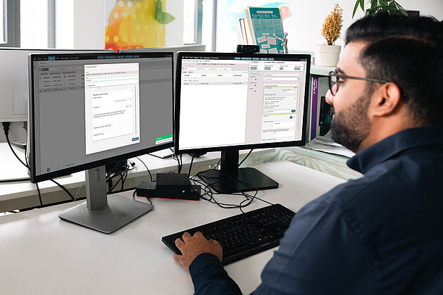
Centralized Management for Clinical Trial Communications and Workflows
Requests for radiological clinical trial evaluations frequently rely on fragmented methods such as emails, phone calls, and spreadsheets. This…
Read more