Funded by the National Institute of Health Research (NIHR) and sponsored by the Imperial College London, the MROC study boasts impressive figures: 645 women with suspected or confirmed ovarian cancer (over 400 recruited at the time of writing), 18 patient recruitment sites across UK and 4 additional external review sites, 22 CRFs per patient, approx. 50 MRI readers and 40 CT readers, as well as several research nurses and surgeons, a histopathologist and a medical oncologist at each site, and a fantastic clinical trial team.
We talked to Prof. Andrea Rockall, Chief Investigator of the MROC study, about her experiences.
What was your main motivation to start such a challenging project?
NICE (National Institute for Health and Care Excellence, UK) published a large evidence document examining the literature on management of women suspected of having ovarian cancer in order to answer the question: What should we be doing to optimize patient care if ovarian cancer is suspected? They identified a lack of high-quality evidence on the role of imaging in accurately determining the best initial treatment. The decision to undertake initial surgery or neoadjuvant chemotherapy is difficult. Some patients undergo ‘open and close’ surgery if the disease is too extensive. Conversely, many women with suspected malignant ovarian mass are ultimately found to have a benign mass and undergo over-extensive or unnecessary surgery, with important implications related to morbidity and fertility. NICE advised the NIHR to commission research to evaluate the role of CT versus MRI (or CT and MRI combined) in staging and determining the optimal treatment if ovarian cancer is suspected. Our proposed study was awarded the grant.
What were your biggest challenges and how did mint help to overcome them?
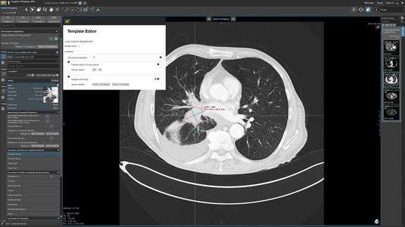
Where do I even start? We have faced numerous challenges from strong study design to meet the commissioning brief, site set up, training of radiographers and radiologists, imaging QA, anonymization and transfer, ensuring blinded sequential imaging review and sequential capture of treatment plans based on the standard of care CT followed by the MRI critical findings and later – treatment plan decisions without knowledge of the CT findings! What I was sure about was that I wanted to develop an electronic CRF that captured both the patient information, imaging CRF with the images, and all the treatment plans and outcomes. We put this job specification out to tender and mint was clearly the closest to being able to deliver the vision.
We have been able to carefully design the CRFs– posing many new challenges to the Mint team. Together we worked tirelessly to develop and support an entire bespoke software platform to host the MROC study, from patient registration to the end of study. The data then feeds into the statistics package.
We have developed a mint training database to teach site staff, including research nurses, radiologists, surgeons and histopathologists. We have expert MRI readers in France, Belgium, and Austria assisting with the blinded reads if local sites need assistance, so we needed on-line training.
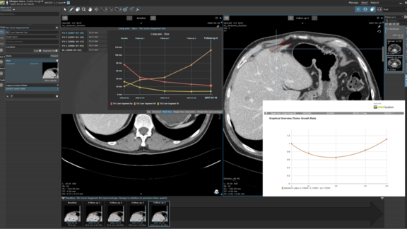
Co-ordinating CT and multiparametric MRI images, blinded double reads, treatment plans, surgical and histopathology findings and outcomes has transformed the way we can deliver image-based clinical trials and bio-banking of study data.
If you were to set up the study now, is there anything you would do differently with the benefit of hindsight?
Certainly. I have made many mistakes along the way. Clear CRF terminology is critical: what I think is a straightforward question can be ambiguous to others. Some of the CRF flow did not quite work correctly for each patient scenario. I would have done more extensive testing of the CRFs prior to study start. We undertook an initial internal pilot in order to evaluate all the processes and we adjusted. mint helped with CRF improvements in order to reduce the variation in responses and reduce data queries. We have now completed our final CRF update and hopefully most readers are comfortable with the CRFs.
How would clinicians and patients benefit from the trial results?
We hope that MROC will improve the radiological assessment of the extent of the disease compared with the current standard of CT scans. This information will potentially help clinicians in multidisciplinary meetings to select a personalized treatment pathway for patients.
For trial summary please visit ISRCTN's website.