Distinguishing between enchondroma, a benign tumor, and low-grade chondrosarcoma, which is a low-aggressivity malignancy, is a frequent challenge, as the lesions are both histologically and radiographically very similar. Given the fact that enchondroma only requires regular follow-up but chondrosarcoma needs surgical resection, differentiating between them becomes extremely important when making a decision on treatment strategies.
Dr. Catharina S. Lisson and colleagues from several departments at the University of Ulm pointed out that MRI-based texture analysis may assist in differentiating these two entities [1]. “The purpose of our study was to identify texture parameters that have the potential to act as non-invasive imaging biomarkers for characterization of chondrosarcoma,” they explained.
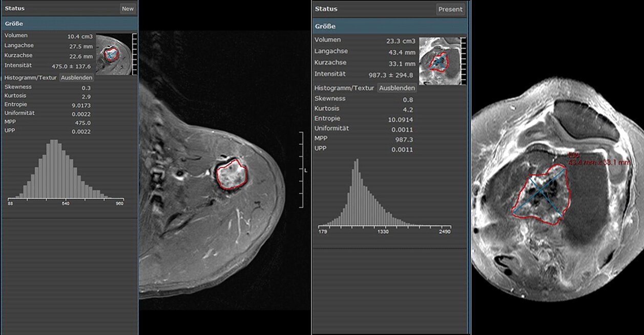
Twenty-two patients were retrospectively evaluated: 11 patients with low-grade chondrosarcoma and 11 patients with enchondroma. Texture analysis was performed in four MRI sequences using mint Lesion. Kurtosis, entropy, skewness, mean of positive pixels (MPP), and uniformity of positive pixel distribution (UPP) were obtained and correlated with histopathology.
According to the results, significant differences were found in four of 20 texture parameters with regard to the different MRI sequences (p < 0.01). In contrast-enhanced T1, the area under the curve (AUC) values to discriminate chondrosarcoma from enchondroma were 0.876 for kurtosis and 0.826 for skewness. In non-contrast T1, the values were 0.851 for entropy and 0.822 for UPP. The highest discriminatory power had kurtosis in contrast-enhanced T1w, with a high sensitivity (82%), specificity (91%), and accuracy (86%).
“Despite encouraging results of emerging studies that show its eligibility in differentiating between tumor types and grading, response monitoring, and outcome prediction, texture analysis has not yet found its way into routine clinical practice,” Dr. Lisson said. “We do hope that further studies with a larger patient population will confirm our findings and thus will lead to a change of the current indication for biopsy and resection of bone lesions.”
[1] Lisson, C.S., Lisson, C.G., Flosdorf, K. et al. Eur Radiol (2018) 28: 468. https://doi.org/10.1007/s00330-017-5014-6