Patients rely on their providers for high-quality care that encompasses the most up-to-date recommendations for diagnosis and therapy. Integrated diagnostics has the potential to pave the way for new diagnostic and therapeutical approaches that, in turn, may significantly improve the patient experience, optimize the patient journey and facilitate better patient outcomes. Mint Medical is committed to supporting healthcare providers focused on bringing more precise and personalized care to their patients by enabling them to combine data from different disciplines and sources. For example, a multidisciplinary approach to diagnosis and therapy is essential in Prostate Cancer – one of the most common cancers amongst men and a leading cause of cancer-related deaths. mint Lesion™ enables healthcare providers to take this multidisciplinary approach to the diagnosis and treatment of prostate cancer.
Screening and Staging
PI-RADS v2.1 reporting in mint Lesion™ is based on the most up-to-date prostate imaging guidelines. We keep enhancing our PI-RADS template to make the read process and reporting simpler, faster, and more consistent for the users.
For whole-body staging of prostate cancer based on PSMA–PET imaging, mint Lesion™ supports guided structured reporting with the ePROMISE template*. The template includes an automated miTNM classification [1], an anatomical region-based hottest lesion determination, as well as other benefits of the standardized mint Lesion™ templates.
Targeted Biopsy
To support targeted biopsy, a mint Lesion™ user can export DICOM Segmentation Objects directly into a guided biopsy device operated by a urologist. This mint Lesion™ feature enables the digital collaboration between radiologists and urologists, simplifying the workflow, saving time, and ensuring high accuracy of the biopsy.
Therapy Response Assessment - Whole Body Bone MRI
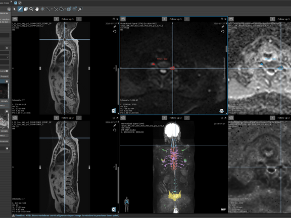
Our AI-powered Bone Disease Assessment template supports the quantification of bone metastases on whole-body MRI in patients with prostate cancer*. In addition to automated quantification of bone disease, the software delivers critical findings in an automatically generated structured report, reducing the turnaround time and streamlining the communication between stakeholders in the clinical pathway.
*not yet commercially available
[1] Prostate Cancer Molecular Imaging Standardized Evaluation (PROMISE): Proposed miTNM Classification for the Interpretation of PSMA-Ligand PET/CT, Matthias Eiber, Ken Herrmann, Jeremie Calais, Boris Hadaschik, Frederik L. Giesel, Markus Hartenbach, Thomas Hope, Robert Reiter, Tobias Maurer, Wolfgang A. Weber and Wolfgang P. Fendler. Journal of Nuclear Medicine March 2018, 59 (3) 469-478; DOI: doi.org/10.2967/jnumed.117.198119


