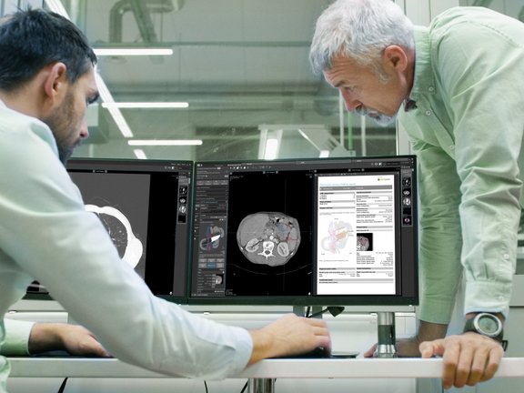
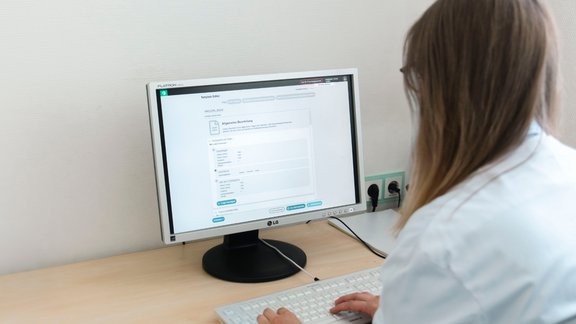
During the daily clinical routine, radiological reports on pancreatic lesions are often written as freeform texts. This causes several problems, in particular the loss of important information, as the level of detail, structure, and terminology used in these reports often varies greatly. In order to ensure an optimal level of patient care, the range of available information must be used in a targeted manner, also in regard to staging and surgical planning. This is facilitated by structured reporting, which enables completeness, reproducibility, and clarity in interdisciplinary communication.
In order to jointly develop structured and quality-assured reading templates for pancreatic lesions in an interdisciplinary consensus meeting, representatives of the German Radiological Society (DRG) met with members of other expert associations, working groups as well as with other radiologists, oncologists, and surgeons. Dr. Matthias Baumhauer, CEO of Mint Medical GmbH, also participated in this meeting. In addition, members of the DRG and the German Society for General and Visceral Surgery (DGAV) were questioned on the topic with the help of an online consensus survey. Subsequently, the experts reviewed all results and compiled them into a manuscript. This acts as a basis for the current reading templates for solid and cystic pancreatic lesions published by the DRG.
Shortly after completion, these templates were also implemented in our software mint Lesion™. They are regularly reviewed and revised by experts, just like the DRG's templates. With mint Lesion™, radiologists can seamlessly integrate all the advantages of structured reporting into their clinical routine. The platform acts as a cognitive assistant to the user and enables the clear and complete preparation of all relevant data. Thus, no information is lost in interdisciplinary communication, which is crucial for optimal clinical care.
Click the here to read the full paper on structured reporting of solid and cystic pancreatic lesions.

Structured Reporting of Solid and Cystic Pancreatic Lesions in CT and MRI: Consensus-Based Reporting Templates of the German Radiological Society (DRG)
Related Resources
Related Resources

RACOON SAGA: Interdisciplinary Research for Better Therapy Decisions in Sarcoma Treatment
Sarcomas are rare tumors that pose particular challenges to both medicine and research. Their heterogeneity makes precise diagnostics difficult, which…

The Template Designer in Use: Interview with Dr. Madelaine Hettler
Structured, well-organized data are the foundation of meaningful research. But how can they be captured effectively in practice? Dr. Madelaine…

Research Insights from Dr. Madelaine Hettler: The Template Designer in Use
Structured and well-organized data are essential building blocks for successful research and clinical projects. The Template Designer provides a tool…