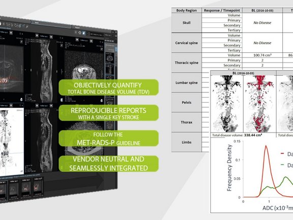
Prostate cancer is the second most common cancer among men, and there are now effective treatments for the disease. New therapies available on the market for treating prostate-cancer metastasis can improve symptoms and prolong life. However, these drugs are often cost-prohibitive. Hence, in order to reduce costs, physicians need to be able to promptly assess the efficacy of therapies, so that ineffective treatments can be quickly terminated and patients switched to a new treatment.
Current imaging modalities for assessing treatment response of bone disease in patients with advanced prostate cancer are unreliable. Standard of care blood tests are inaccurate and alongside conventional imaging such as the bone scan and computed tomography (CT) have limitations when it comes to assessing and quantifying treatment response. Whole body diffusion-weighted (WBDW) MRI demonstrates high sensitivity in detecting bone disease from prostate cancer with the benefit of being non-invasive, without ionising radiation or requiring any contrast medium injection.
Continuing the tradition of revolutionizing therapy-response assessments, at the upcoming 104th annual meeting of RSNA, Mint Medical will announce its latest decision support prototype that allows radiologists to quantify total tumor load of bone disease in patients with metastatic prostate cancer within minutes to help management decisions. This tool is developed in cooperation with The Royal Marsden NHS Foundation Trust and The Institute of Cancer Research, London, with funding from the National Institute for Health Research (NIHR) and support from the NIHR Biomedical Research Centre at The Royal Marsden NHS Foundation Trust and The Institute of Cancer Research (ICR). Prof. Dow-Mu Koh MD, MRCP, FRCR, Consultant Radiologist in Functional Imaging at The Royal Marsden and Professor in Functional Cancer Imaging at the ICR, who is leading the project, said:
“With Mint Medical, we are developing software that allows us to detect and outline regions of disease from WBDWI and thus estimate tumour volume and ADC very quickly. We hope this methodology will provide an easy-to-use approach for reporting response of metastatic prostate disease to systemic treatments, adopting recently guidelines for standardised reporting of WBDWI (MET-RADS-P) [1] in prostate cancer.”
In addition to automated quantification of bone disease, mint Lesion automatically prepares a structured report for radiologists with critical findings, further reducing the turn-around-time, increasing the quality of the reports and streamlining the communication to urologists.
“Due to their intelligently designed user workflows with a structured-data backend architecture, mint Lesion is able to obtain these measurements in a significantly reduced time, whereas other available commercial methods would often take from up to 30 minutes and be restricted to specific vendors,” continues Prof. Koh.
“We have well-defined short-term goals directly linked to advancing decision-support tools in diagnostic imaging,” says Dr. Matthias Baumhauer, co-founder and CEO of Mint Medical. “We don’t focus on hypothetical tinkering in our AI laboratory. On the contrary, we are laser-focused on building tools with our clinical partners that will make a difference in the immediate radiologists‘ workflow and empower them with insights to make better therapy response decisions today. In addition, mint Lesion supports all imaging modalities vendors, making it available to the entire global diagnostic imaging community,” continues Baumhauer.


