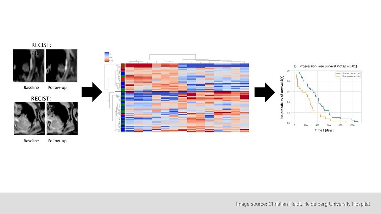
In this prospective study conducted by Heidelberg University Hospital, researchers investigated whether changes in radiomic features from diffusion-weighted MRIs (DWI) could provide early indications of treatment success in patients with advanced lung adenocarcinoma. A total of 144 patients were analyzed, all of whom were treated either with tyrosine kinase inhibitors (TKI) or platinum-based chemotherapy (PBC).
The aim of the study was to use delta-radiomics features (DRFs)—which represent changes in radiomic features over time—to gain early, tissue-level insights into treatment success. Patients were examined with DWI-MRI both before treatment and at several points after therapy began. Particular attention was given to changes in Apparent Diffusion Coefficient (ADC) maps, which offer insights into tissue changes.
“Although frequently used in chest CT imaging, delta-radiomics applied to functional imaging remains understudied. Previously, functional imaging, such as perfusion-MR or diffusion-weighted MR imaging (DWI), has shown earlier detection of tissue changes compared to morphological imaging.”
The study's findings can be summarized as follows:
- As early as 14 days after the start of treatment, specific radiomic features could be correlated with treatment success and progression-free survival (PFS) of the patients.
- Patients who responded better to treatment showed increased values in certain delta-radiomics features (DRFs), which captured the heterogeneity of the tumor regions, indicating an increase in diffusivity within the tumor.
- In contrast, patients with poorer responses showed an increase in features that indicated a more homogeneous tissue structure, suggesting a stable or increasing tumor tissue density.
The study highlights that the analysis of delta-radiomics features can provide valuable information about the course of therapy after a short period of time. This method could potentially help to adjust treatments more quickly in the future by identifying non-responding patients early and considering alternative treatment options.
Lung cancer is the leading cause of cancer-related deaths worldwide, and after prostate and breast cancer, it is the third most common cancer diagnosis in both sexes. Since early stages are often asymptomatic, the disease is usually diagnosed at an advanced stage, worsening the prognosis and limiting treatment options.
The use of diffusion-weighted MRI and delta-radiomics analyses offers a promising, radiation-free alternative to conventional imaging for monitoring lung cancer treatments. It enables the early prediction of treatment success, potentially speeding up therapy decisions and adjustments. However, further studies are needed to confirm the effectiveness of this method in larger patient cohorts and to determine the optimal time frame for assessing treatment response.
Read the original publication here: https://insightsimaging.springeropen.com/articles/10.1186/s13244-024-01787-5
Heidt, Christian M. et al. 2024. “Delta-radiomics features of ADC maps as early predictors of treatment response in lung cancer.” Insights Into Imaging 15.218.