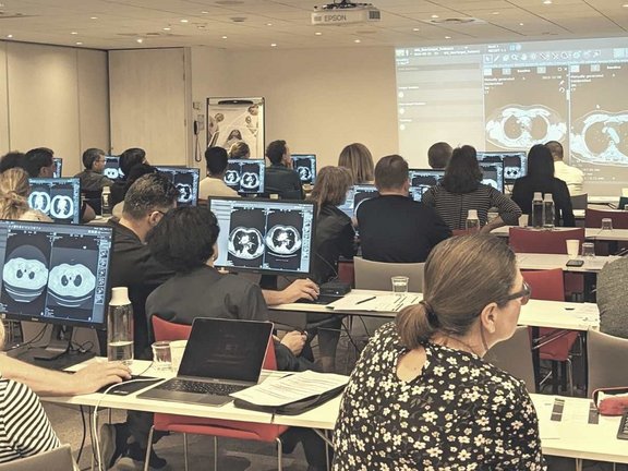
Last week Mint Medical supported an ESOI-EORTC joint hands-on course on imaging in assessing response to cancer therapy - a yearly event that took place in Bordeaux, France.
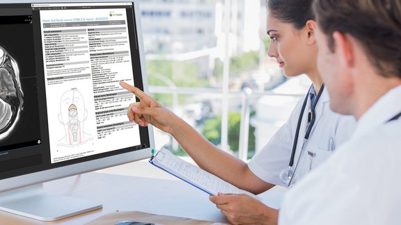
During this course, the participants had an opportunity to learn the principles of imaging-based tumor response assessment, review the most common response criteria, dive into the principles of structured oncological reporting and gain relevant knowledge about clinical trials and trial methodology. After each lecture, the participants had the chance to evaluate and analyze relevant real-life cases encountered in the clinical routine or within clinical trials, using mint Lesion™ as dedicated software.
We spoke with one of the organizers and lecturers of the workshop - Professor Laure Fournier, from Hôpital Européen Georges Pompidou, Paris Cité University.
MM: What was the idea behind this workshop?
LF: "Since EORTC is really implicated in clinical trials, where oncologic imaging plays a key role, we wanted to train the European radiology community to be proficient with imaging criteria for assessment of treatment response. The best way to ensure such proficiency would be through hands-on training of the radiologists involved in oncologic imaging. ESOI was experienced at organizing similar trainings, so we decided to organize our first joint workshop in 2016, which has become a successful yearly event."
MM: What is the primary purpose and value of such workshops?
LF: "The main purpose of these workshops is to foster consistent application and interpretation of the criteria across Europe and beyond, as well as to bring the whole radiology community dealing with cancer imaging to the expected level of quality. The opportunity to assess and then discuss challenging cases is the most valuable aspect of the course. It's not just about theoretical lectures. It's about getting into each case's details and learning from them. This hands-on experience is what makes the most significant difference for the participants. In addition, they really appreciate the opportunity to interact and compare experiences with colleagues from all over the world in a casual, friendly atmosphere. That's why the workshop has been fully booked every single time."
MM: Who can benefit from this workshop?
LF: "Theoretically, any radiologist can benefit, but more specifically, those who are regularly involved in oncologic imaging activity. The ideal candidates are the radiologists who have basic knowledge of the response criteria and have already started applying them but are not fully comfortable with them. This course will allow them to deepen their knowledge and pick up more complicated aspects."
MM: Does the content of the workshop change every year?
LF: "While the essential parts, focusing on the response criteria, stay unchanged, some of the lectures underlining certain aspects of cancer imaging evaluation vary from year to year. Every time there are new horizon lectures, which are innovative and offer an outlook into the future."
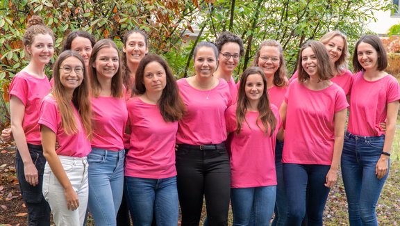
For us, in Mint Medical, this workshop was again a great experience. We want to thank Melvin D'Anastasi from Mater Dei Hospital in Malta, Michèle Kind from the Institut Bergonié in Bordeaux, and everyone else who organized this year's workshop. Looking forward to supporting this amazing event next year, wherever this may take us.


