This study explores the use of deep learning (DL) to optimize MRI protocols for glioblastoma patients. Glioblastomas, known for being the most prevalent and most aggressive malignant brain tumors in adults, require frequent and detailed monitoring through MRI - a process that can be particularly challenging for critically ill patients due to the lengthy scan times and potential for motion artifacts. This study thus focuses on assessing the effectiveness of a DL-optimized MRI protocol that aims to reduce scan time while improving image quality.
Conventional approaches, such as parallel acquisition techniques (PATs) and compressed sensing (CS), have been utilized to shorten examination times and address related challenges. However, these methods come with limitations: PATs can lead to a decrease in the signal-to-noise ratio proportional to the square-root of the PAT factor, while CS may produce images that are overly smooth and lack realistic detail.
The researchers underline the potential of DL to optimize MRI protocols: “Integrating DL reconstruction (DLR) techniques into medical imaging offers the potential to improve diagnostic accuracy and efficiency in glioma patients. Nevertheless, further research is warranted to systematically investigate the quality of DL-generated images and optimize their practical application in routine clinical settings.”
The study involved 33 patients with histologically confirmed glioblastoma, who underwent brain tumor imaging on a 3-Tesla MRI scanner. Two experienced neuroradiologists conducted independent assessments of the image datasets, focusing on subjective image quality, diagnostic confidence, tumor visibility, noise levels, artifacts, and sharpness. Additionally, they measured tumor volume based on the Response Assessment in Neuro-Oncology (RANO) 2.0 criteria, comparing the results between both imaging techniques and evaluating various clinical-pathological parameters.
All in all, key findings from the study include:
- Time Efficiency: The DL-optimized MRI sequences reduced scan time by an average of 30% without compromising the quality of the scans.
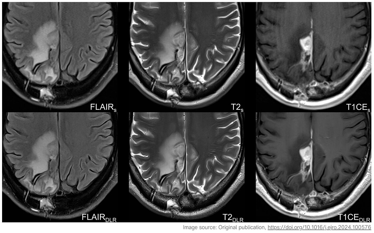
- Enhanced Image Quality: The DL-enhanced MRI sequences demonstrated superior image quality, including sharper images, better tumor visibility, and reduced noise levels, improving overall diagnostic capabilities. These improvements are crucial for maintaining diagnostic confidence and accuracy.
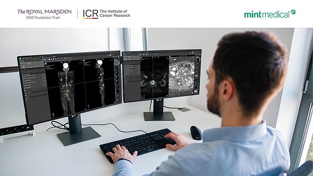
- Maintained Diagnostic Accuracy: Using mint Lesion™ software, the study confirmed that the DL-optimized protocol did not negatively impact tumor volume measurements or response assessments, ensuring consistency with standard neuro-oncology practices. This was validated by the RANO 2.0 criteria.
- Practical Benefits: The faster scan times and improved image quality are particularly beneficial for patients who struggle to remain still during lengthy MRI procedures, reducing the need for repeat scans and minimizing motion artifacts.
This study underscores the potential of DL-optimized MRI protocols to enhance the efficiency and quality of glioblastoma imaging, offering a promising approach for clinicians and patients alike.
Read the original publication here: https://www.mdpi.com/2072-6694/16/10/1827
Gohla, Georg, Till-Karsten Hauser, Paula Bombach, et al. 2024. „Speeding Up and Improving Image Quality in Glioblastoma MRI Protocol by Deep Learning Image Reconstruction.” Cancers 16.10: 1827.