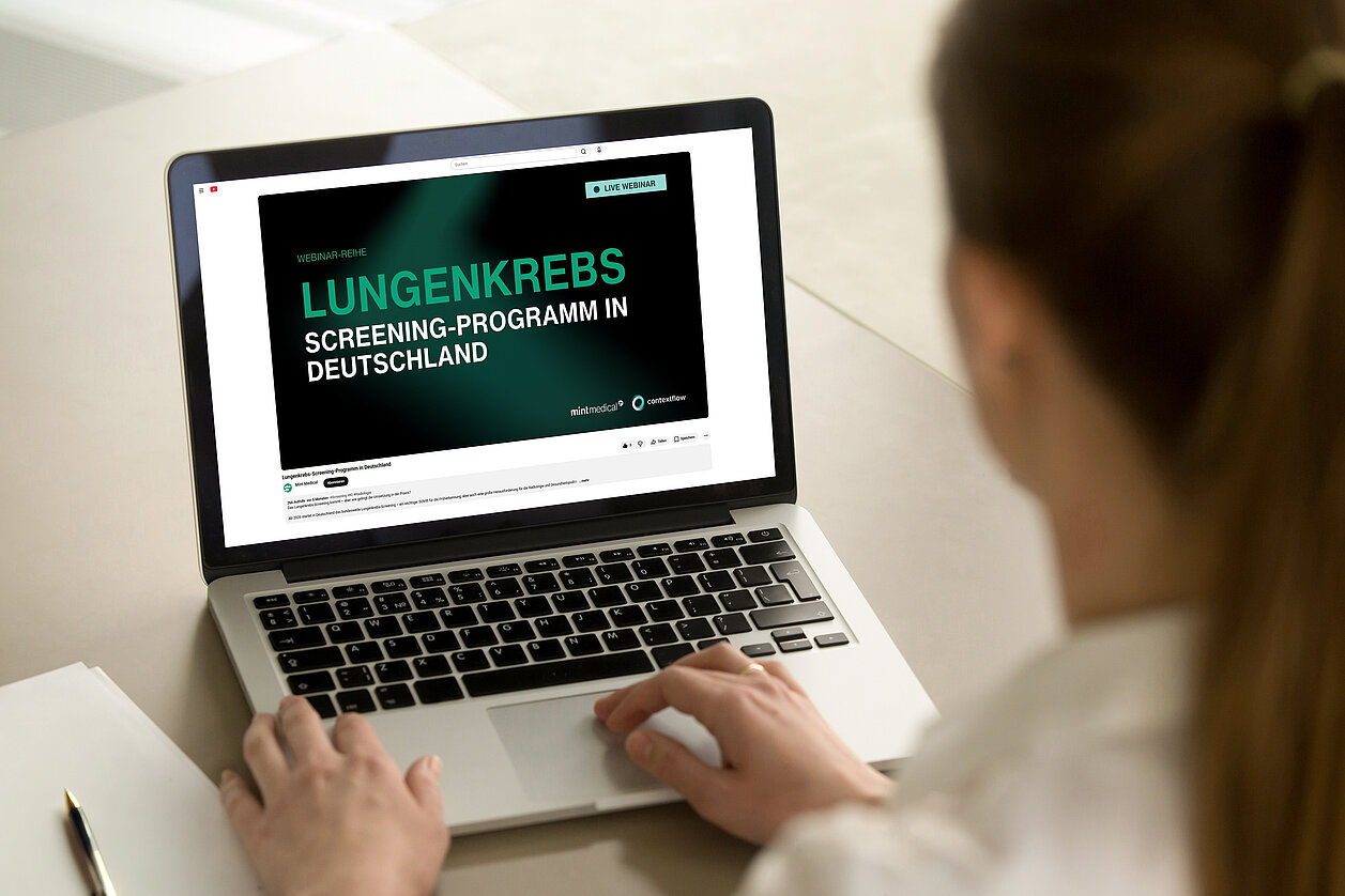
Webinar Series: Lung Cancer Screening in Germany - From Evidence to Implementation
The program is based on strong scientific evidence, including large international trials such as NLST and NELSON, which have demonstrated a significant reduction in mortality through early detection. [1]
However, translating these findings into clinical practice requires more than technology alone. It involves clearly defined workflows, technical standards, and structured data management across multiple stakeholders.
Together with contextflow, we have launched a webinar series to explore different aspects of lung cancer screening in Germany. On this page, we bring together the first three sessions—providing all key insights in one place.
Want to understand how lung cancer screening can be implemented efficiently and at scale?

Webinar Series: Insights into Lung Cancer Screening in Germany
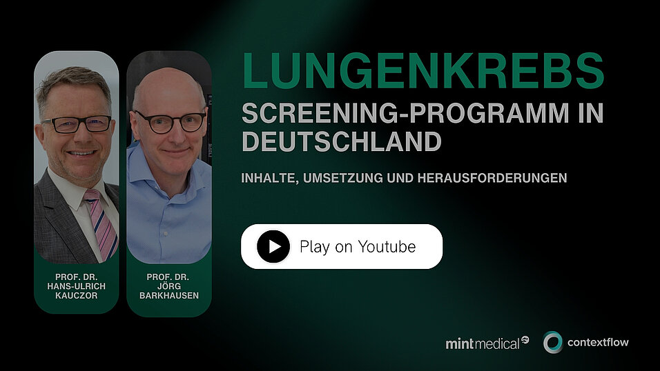
#1: Content, Implementation, and Challenges
This session provides a comprehensive introduction to the medical rationale and regulatory framework of lung cancer screening in Germany.
Experts highlight the importance of early detection through LDCT, enabling a shift from late-stage, often incurable diagnoses to earlier, treatable stages. At the same time, the discussion addresses key challenges such as radiation exposure, false positives, and overdiagnosis.
The webinar also outlines the current legal status in Germany, including technical requirements for imaging systems and the mandatory use of AI-based software for nodule detection. Reimbursement for statutory health insurance is expected to follow. [At the time of the webinar, reimbursement for statutory health insurance had not yet been finalized. This information has since been updated: read more here.]
Speakers: Professor Hans-Ulrich Kauczor (University of Heidelberg) and Professor Jörg Barkhausen (University of Lübeck), prominent thought leaders in the field of lung cancer screening, as they have been instrumental in preparing the medical and legal frameworks for the program's implementation in Germany and across Europe.
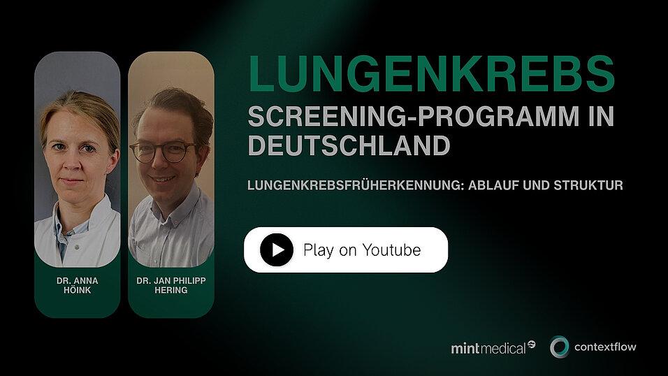
#2: Workflow and Structure
The second webinar focuses on the practical implementation of the screening program and the requirements for participating institutions.
It covers eligibility criteria for participants, certification requirements for radiologists, and the structured workflow involving referring physicians, first readers, and specialized second readers.
In addition, the session highlights technical requirements for CT scanners and emphasizes the importance of digital platforms to manage data exchange and coordination across all stakeholders involved in the screening process.
Speakers: Dr. Anna Höink (University Hospital OWL) and Dr. Jan-Philip Herring (Klinikum Ibbenbüren), both members of the "Lung Cancer Early Detection Taskforce".
Interested in how structured workflows and interoperable systems can support these processes? Get in touch with our team.
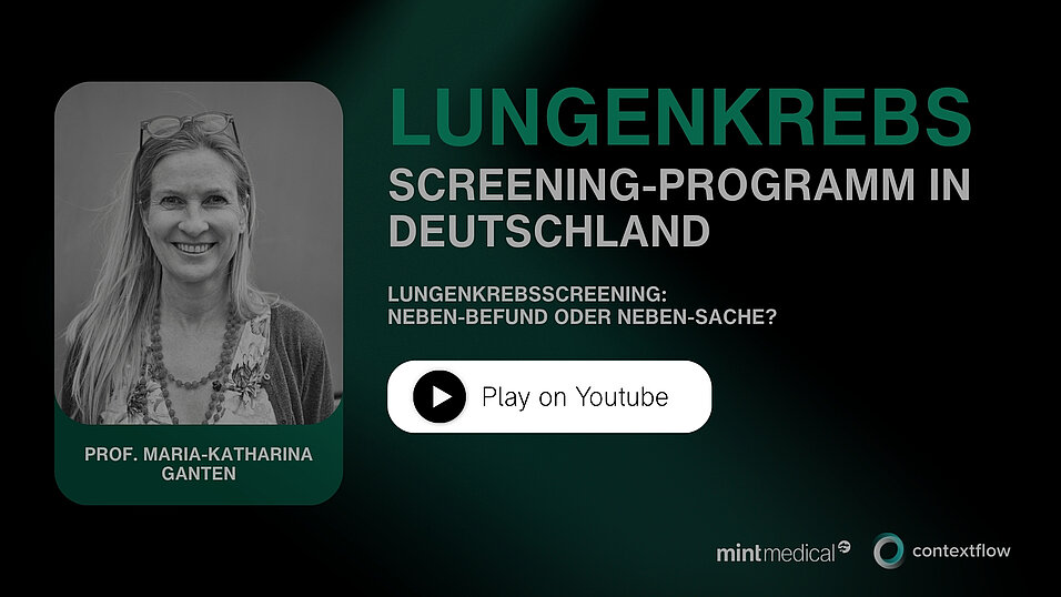
#3: Incidental Findings – Managing Complexity
The third webinar addresses one of the most challenging aspects of lung cancer screening: the management of incidental findings.
Based on international guidelines and practical experience, the session demonstrates how to balance clinical relevance with the need to avoid unnecessary patient anxiety. The principle of “keep it simple” is central—only findings that require clinical action should be highlighted.
Examples of actionable findings include aortic aneurysms, significant emphysema, or suspicious nodules in other organs. At the same time, minor or stable findings should be documented without triggering unnecessary follow-up.
The webinar also highlights how standardized reporting approaches, as seen in established programs such as in the UK, can support consistent and efficient decision-making.
Speaker: Professor Maria-Katharina Ganten, an expert with extensive experience in the UK’s lung screening program
From Evidence to Implementation
Lung cancer screening in Germany is moving from concept to reality. Its success will depend on structured workflows, reliable data management, and seamless collaboration across institutions.
Mint Medical supports these requirements with software designed for standardized data collection, structured reporting, and interoperability—helping to enable scalable and efficient screening programmes.
Get in touch to learn how mint Lesion can support your lung cancer screening workflows.

[1] Charlotte Poon et al. 2022. Should we screen for lung cancer? A 10-country analysis identifying key decision-making factors.” Health Policy, 126.9: 879-888. https://doi.org/10.1016/j.healthpol.2022.06.003.
Related Resources

The ICR and The Royal Marsden work with Mint Medical to integrate AI-powered software into cancer treatment

RadMag Special Edition: Onur Özek on the Challenges of Lung Cancer Screening in Germany