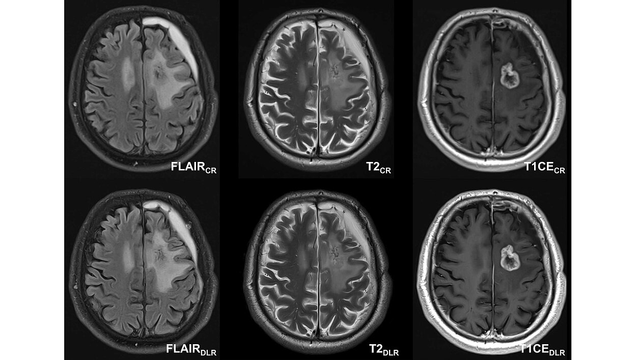
A recent study conducted by the University Hospital of Tübingen investigated the potential of deep learning reconstruction (DLR) in magnetic resonance imaging (MRI) for IDH-mutant gliomas compared to conventional imaging (CR). The study was carried out by an interdisciplinary team of neuroradiologists, neurosurgeons, neuro-oncologists, and radiation oncologists. The objective was to evaluate the reduction in acquisition time, image quality, and diagnostic confidence of DLR images within a comprehensive diagnostic MRI protocol, including contrast-enhanced T1-weighted, T2-weighted turbo spin echo (TSE), and FLAIR sequences.
The researchers hypothesize that DLR would provide superior quantitative and qualitative image quality, benefiting all specialists involved in the diagnosis and treatment of gliomas, as well as the patients themselves.
The key findings of the study can be summarized as follows:
Time Savings: DLR significantly reduced MRI acquisition times across all sequences:
- 35.8% for T1-weighted contrast-enhanced imaging
- 26.9% for FLAIR-weighted imaging
- 26.1% for T2-weighted imaging
Image Quality: DLR consistently received higher qualitative image quality ratings from all raters across all sequences. Tumor conspicuity with DLR was non-inferior to CR images.
Multidisciplinary Preference: The majority of raters preferred DLR images over CR:
- 84% of neuroradiologists
- 100% of neurosurgeons
- 92% of neuro-oncologists
- 84% of radiation oncologists
Enhanced Metrics: Quantitative analysis demonstrated significant improvements in signal-to-noise ratio (SNR) and contrast-to-noise ratio (CNR) with DLR. Measurements of lesion size showed no significant differences between CR and DLR sequences according to the criteria for tumor assessment and treatment response (RANO 2.0 criteria).
The study concluded that DLR is clinically feasible for MR imaging of IDH-mutant gliomas. It offers significant time savings (reducing acquisition time by an average of 29.6%) without compromising image quality or diagnostic accuracy. The strong preference for DLR among the multidisciplinary team highlights its potential to streamline neuro-oncological workflows and improve decision-making.
With these findings, DLR represents a promising advancement in MRI, enabling faster imaging with exceptional clarity to support the effective management of gliomas.
Read the original publication here.
Ruff, Christer et al. 2024. „Multidisciplinary quantitative and qualitative assessment of IDH-mutant gliomas with full diagnostic deep learning image reconstruction.” European Journal of Radiology Open, Volume 13, December 2024, 100617, doi.org/10.1016/j.ejro.2024.100617.