
Radiologic Recurrence Criteria for Adjuvant Clinical Trials in Bladder Cancer
Response criteria in oncology imaging rest on a simple premise: there is a tumor, you measure it, and you watch what treatment does to it. RECIST works because there is something on the scan to follow.
The adjuvant setting inverts that. After radical surgery the patient is — by definition — free of measurable disease, so the question is no longer how much smaller? but is anything coming back? Conventional criteria were never designed to say when a finding on a surveillance scan counts as unequivocal cancer. The criteria developed by Apolo et al. for adjuvant clinical trials in bladder cancer close that gap.
What are the Radiologic Recurrence Criteria for Adjuvant Trials?
The radiologic recurrence criteria for adjuvant trials referenced in Apolo et al. are a consensus framework harmonizing eligibility requirements, the definition of "no evidence of disease," and the radiologic definition of disease recurrence in adjuvant clinical trials — originally for muscle-invasive bladder cancer (MIBC). They emerged from a public workshop convened by the US Food and Drug Administration and the National Cancer Institute, supported by the Society of Urologic Oncology, on 28 November 2017 at the National Institutes of Health in Bethesda, Maryland, with medical and urologic oncologists, radiologists, biostatisticians and patient advocates. Andrea B. Apolo and colleagues published the conclusions in JAMA Oncology in 2019.
The framework is deliberately pragmatic: its authors state plainly that robust data for specific thresholds are still lacking and that the definitions will need adjustment. What the criteria buy is not biological certainty but consistency — if every site applies the same rule to the same equivocal nodule, the resulting disease-free survival curve means something.
At screening: is the patient truly disease-free?
Where no prior imaging exists, a 1.0 cm decision threshold applies — long axis for non-nodal lesions, short axis for lymph nodes:
- An equivocal lesion below 1.0 cm is assumed benign.
- A lesion of 1.0 cm or more is treated as suspicious. Absent an alternative clinical explanation, it warrants targeted workup, a repeat examination, or biopsy before enrollment.
The cutoff reflects both the radiologic consensus on the size at which a lesion becomes reasonably sensitive and specific for cancer, and the size needed to characterize it reliably on CT.
During surveillance: what counts as unequivocal recurrence?
Designed with RECIST principles in mind and underpinned by Quantitative Imaging Biomarkers Alliance (QIBA) standards, any of the following counts as unequivocal recurrence once a non-malignant explanation has been excluded:
- A new lesion of 1.0 cm or more absent at the initial evaluation.
- A pre-existing lesion below 1.0 cm with more than 50% growth across two consecutive examinations plus an absolute increase of at least 5 mm.
- A pre-existing lesion of 1.0 cm or more with more than 50% growth on a single examination.
- Multifocal sub-centimeter lesions whose distribution or radiologic/metabolic features are pathognomonic for metastatic disease.
The two-examination requirement is the central safeguard: calling recurrence on one measurement of a 6 mm nodule would badly overestimate events, whereas sustained growth of this magnitude clearly outpaces metabolically indolent processes — specific enough to stand in for histology when biopsy is unavailable.
Anatomic nuance and acquisition discipline
- Lymph nodes: short axis; 1.0–1.5 cm is suspicious but rarely convincing on imaging alone, ≥1.5 cm highly suspicious, especially when growing across scans.
- Liver: any new enhancing lesion is suspicious, even below 1.0 cm.
- Bone: a new lytic or sclerotic lesion qualifies; CT features are often non-specific, so unclear lesions need radiography, MRI or nuclear medicine.
- Pleural effusion and ascites: no modality separates benign from malignant fluid — sample for cytology where safe and feasible.
- Resection bed: image at least four weeks post-surgery, once inflammation has resolved.
- Technique: slice display alone changes detection rates and measured diameters, so acquisition, display and interpretation should follow QIBA guidance. Contrast-enhanced CT of chest, abdomen and pelvis is the modality of choice, within four weeks before enrollment; with renal insufficiency, non-contrast chest CT plus gadolinium MRI of abdomen and pelvis substitutes.
The date of recurrence: a deliberate open question
Backdating to the scan of first appearance is temporally accurate but inconsistent — the anchor may be a finding that never met trial criteria. The date the prespecified criteria are met is reproducible but lags true onset. Neither was mandated: the protocol must state its choice and apply it uniformly. Where a radiologic and a histopathologic date both exist, the radiologic date is preferred, since biopsies are usually prompted by imaging.
Harmonized eligibility
The same publication tackles the heterogeneity created when chemotherapy-era entry criteria are carried into trials of checkpoint inhibitors and other novel agents:
- Variant histology eligible alongside predominant urothelial carcinoma; pure non-urothelial histologies, especially mixed neuroendocrine/small-cell, analyzed separately.
- Neoadjuvant chemotherapy defined as ≥3 cycles of cisplatin-based combination therapy at a planned 70 mg/m² per cycle; below that, patients are stratified as untreated and judged on post-cystectomy stage.
- Muscle-invasive upper-tract urothelial carcinoma included, ideally stratified against lower-tract disease.
- Severe renal insufficiency no barrier to trials of agents that are not renally excreted.
- Microscopic (R1) margins eligible, stratification considered; grossly positive (R2) margins unresolved.
- Bilateral standard pelvic lymph node dissection before enrollment — favored and sufficient for staging.
- Adjuvant therapy started once the patient has recovered, one to four months post-surgery.
- New urothelial tumors as events: all new high-grade upper-tract and all new muscle-invasive bladder tumors count toward disease-free survival; ≤T1 bladder tumors remain unclear; low-grade non-muscle-invasive disease manageable endoscopically may stay on trial. For urethral second primaries, non-muscle-invasive disease manageable endoscopically or by urethrectomy permits continuation, while muscle-invasive recurrence is an event and ends participation.
What this means for the reading workflow
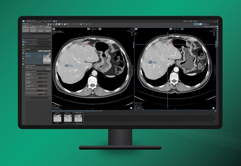
Operationally, the criteria are more demanding than RECIST. A single recurrence call can depend on three comparisons — against baseline, against the previous timepoint and, for small pre-existing lesions, the timepoint before that. Each lesion must be categorized as new, pre-existing-small or pre-existing-large, because the rule differs by category, and absolute and percentage change both have to be right. Done manually across dozens of sites and hundreds of timepoints, this is where reproducibility quietly erodes.
Hence implementing the framework in the reading software itself. In mint Lesion, the radiologic recurrence criteria for adjuvant trials are available as a standardized assessment framework, providing:
- Structured lesion categorization — pre-existing large, pre-existing small and new lesions, with configurable size thresholds, so protocols with different cutoffs remain supported.
- Automated change tracking — absolute and percentage diameter change against baseline, the previous timepoint and, for small pre-existing lesions, two timepoints prior.
- Guided recurrence determination — status derived from the trial's uniform definitions instead of reconstructed by hand.
For sponsors and imaging teams the benefit is structural: consistent disease-status monitoring and reliable data capture across multicenter trials, with the criteria enforced in the workflow rather than left to memory.
Although the publication by Apolo et al. is specifically defined for bladder cancer, the implementation of the criteria in mint Lesion is intended to be applicable to other solid tumor indications assessed in an adjuvant setting.
Caution: The criteria by Apolo et al. are not validated in settings other than adjuvant bladder cancer, and Mint Medical has not validated them for any other use. We recommend performing a validation before applying the criteria in a different setting.
Outlook
The adjuvant criteria are consensus, not settled science — and their authors say so. Protocols may choose different thresholds provided they are applied consistently, and the FDA has recommended continued dialogue during trial design as evidence matures. What the framework already achieves is more modest and more useful: it turns "is this recurrence?" from a matter of local radiologic culture into a question with a prespecified, auditable answer.
Frequently asked questions
What are the criteria by Apolo et al. in oncology imaging?
The criteria by Apolo et al. are a consensus framework for adjuvant clinical trials, published in JAMA Oncology in 2019 after a workshop convened by the US Food and Drug Administration (FDA) and the National Cancer Institute (NCI). They standardize eligibility, the definition of no evidence of disease and the radiologic definition of recurrence. They were initially developed for muscle-invasive bladder cancer (MIBC).
How do the criteria by Apolo et al. differ from RECIST?
RECIST tracks existing target lesions in patients with measurable disease. The criteria by Apolo et al. address the adjuvant setting, where patients have no measurable disease after surgery and the endpoint depends on new lesions. RECIST provides no size or growth rules for such new lesions.
What size does a new lesion need to reach to count as recurrence?
Under the criteria by Apolo et al., a new lesion counts as unequivocal recurrence if it measures 1.0 cm or more, was absent at the initial evaluation and a non-malignant explanation has been excluded. Lymph nodes are measured in short axis, all other lesions in long axis. Smaller lesions can also count as recurrence if they are multifocal and their distribution or radiologic or metabolic features are pathognomonic for metastatic disease.
How is growth of a pre-existing lesion assessed?
For pre-existing lesions, the criteria by Apolo et al. distinguish by size. A lesion below 1.0 cm counts as recurrence if it grows by more than 50% across two consecutive examinations and increases by at least 5 mm in absolute terms. A lesion of 1.0 cm or more counts as recurrence if it grows by more than 50% on a single examination. In both cases, a non-malignant explanation must be excluded.
How is the date of recurrence determined?
The criteria by Apolo et al. describe two options: backdating to the scan on which the finding first appeared, which is temporally accurate but less consistent, or using the date on which the prespecified criteria are met, which is reproducible but lags true onset. Neither option is mandated. The trial protocol must specify one and apply it uniformly.
Do the criteria by Apolo et al. apply only to bladder cancer?
The criteria by Apolo et al. were developed for muscle-invasive bladder cancer and are not validated outside the adjuvant setting in bladder cancer. The implementation in mint Lesion is intended to be applicable to other solid tumor indications in the adjuvant setting as well. Before the criteria are used in any other setting, a validation should be performed.
Are the criteria by Apolo et al. a regulatory requirement?
No. The criteria by Apolo et al. are consensus recommendations from a workshop convened by the FDA and the NCI, not a regulatory requirement. Trial protocols may adapt the thresholds, provided the definitions are prespecified and applied consistently.
Reference
Apolo AB, Milowsky MI, Kim L, et al. Eligibility and Radiologic Assessment in Adjuvant Clinical Trials in Bladder Cancer. JAMA Oncology. 2019;5(12):1790–1798.
Related Resources

Postoperative Brain MRI After Tumor Resection: Reducing Artifacts and Improving Diagnostic Accuracy

One Place to Request, Track, and Document Trial Reads