jump to content jump to footer
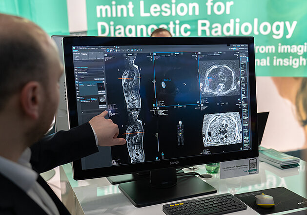
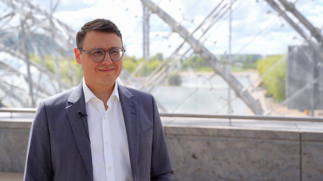
Prof. Dr. Timm Denecke discusses AI-based early detection of liver cancer at RÖKO 2026

Predicting hepatocellular carcinoma (HCC) before it develops: Artificial Intelligence meets MRI

At RÖKO 2026, we spoke with Prof. Dr. Timm Denecke, Director of Radiology at Leipzig University Hospital, about RACOON-MARDER, a research project using artificial intelligence to detect the earliest imaging signs of hepatocellular carcinoma (HCC) in high-risk patients. By training AI on structured MRI data from across Germany's university hospitals, the project aims to identify which patients truly need intensive screening — and catch liver cancer before it becomes cancer. Mint Medical is proud to support this research as a technical network partner.

Read the full interview to find out how AI could reshape liver cancer screening.

Or watch the video interview with Prof. Denecke on Youtube. 
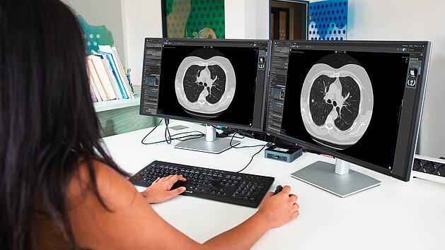
Hospital staff using mint Lesion for interoperable workflows and data management in lung cancer screening
How mint Lesion supports hospitals and screening centers with interoperable infrastructure, AI integration, data management, and scalable workflows for lung cancer screening.
Lung Cancer Screening in Germany: How mint Lesion Supports Hospitals with Infrastructure, Integration, and Scalability
Screening as a Strategic Challenge With the launch of the national lung cancer screening program in 2026, hospitals and screening centers across…
Read more
Radiologist analyzing whole-body MRI scans of prostate cancer bone metastases using AI-assisted tumor load quantification in mint Lesion
How mint Lesion supports radiologists in AI-assisted tumor load quantification for bone metastases in prostate cancer with structured analysis, objective metrics, and longitudinal therapy assessment.
AI-Supported Tumor Load Quantification for Bone Metastasis in Prostate Cancer
To assess treatment response in patients with advanced prostate cancer, radiologists rely on advanced medical imaging. Conventional modalities, such…
Read more
Radiologist analyzing whole-body MRI scans of multiple myeloma using AI-assisted quantification in mint Lesion
How mint Lesion supports radiologists in AI-assisted quantification of bone involvement in multiple myeloma with structured analysis, objective metrics, and longitudinal disease tracking.
AI-Supported Quantification of Bone Involvement in Multiple Myeloma
Radiologists utilize Whole-Body MRI (WB-MRI) as an established imaging method for multiple myeloma staging [1,3]. Because it avoids ionizing…
Read more
scroll-top