jump to content jump to footer
Radiologist using mint Lesion for structured lung cancer screening reporting and AI-assisted lung nodule assessment

Lung Cancer Screening in Germany: How mint Lesion Supports Radiologists in Clinical Practice

A New Era of Early Detection — and New Challenges

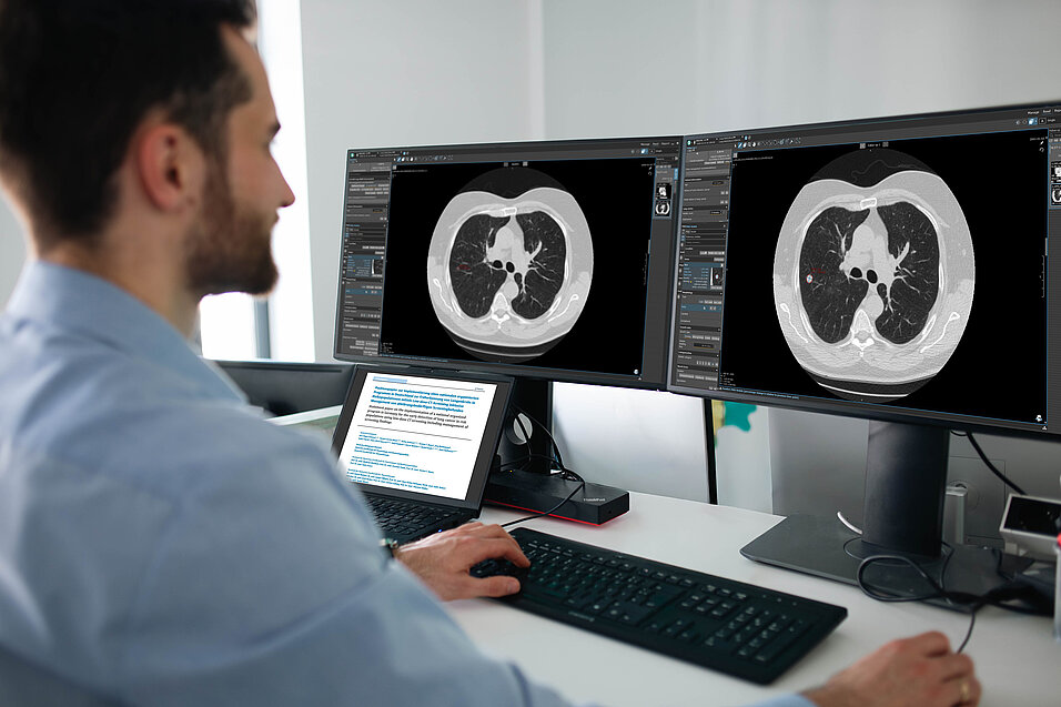
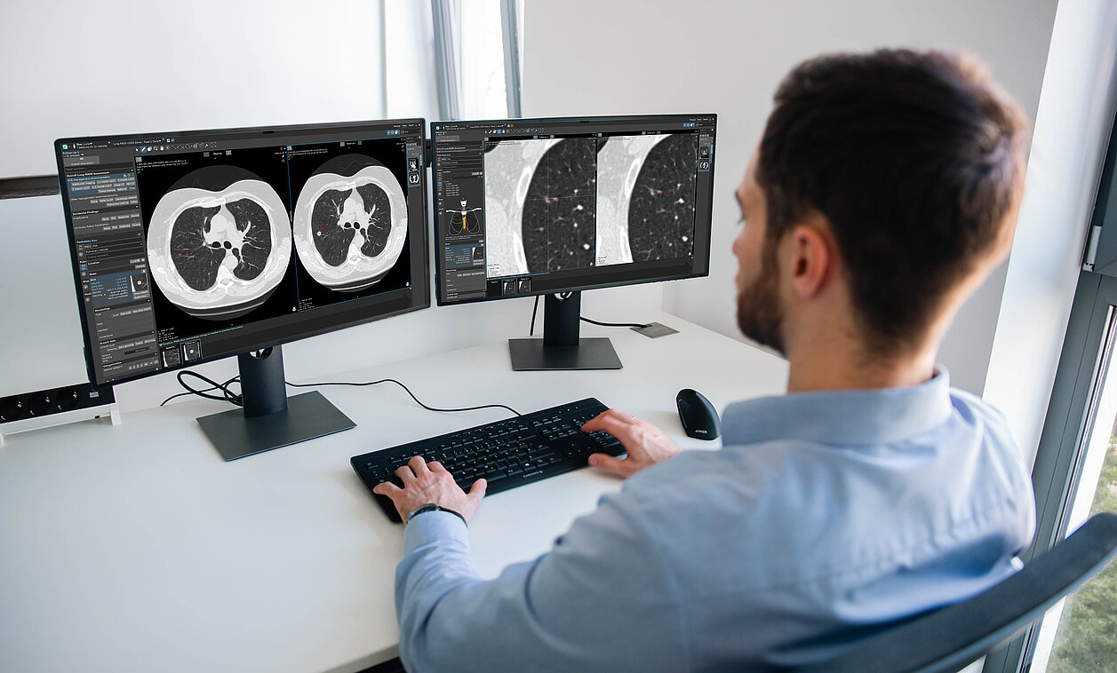
With the introduction of the nationwide lung cancer screening program starting in 2026, radiologists in Germany are entering a new era of early detection. Standardized low-dose CT (LDCT) examinations, clearly defined eligibility criteria, and mandatory second readings are expected to improve diagnostic quality—but they also introduce significantly greater complexity and workload.

Today, lung cancer screening involves far more than image interpretation alone. It requires structured reporting, reliable longitudinal follow-up, close collaboration with second-reading institutions, and strict adherence to clinical guidelines.

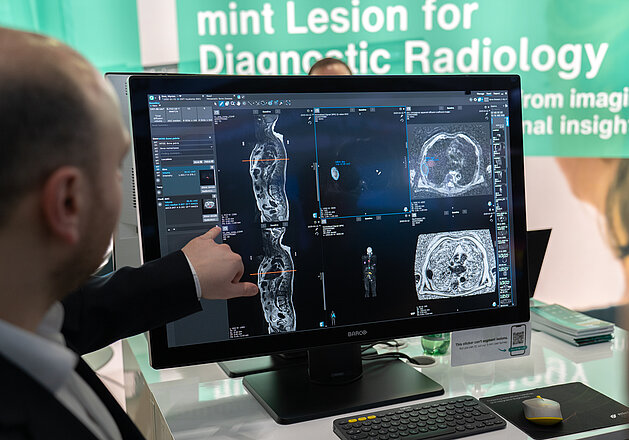
This is exactly where mint Lesion comes in.

Structured Reporting — Consistent and Guideline-Compliant

In lung cancer screening, structured reporting is essential for quality, comparability, and regulatory compliance. Variability in interpretation and inconsistent reporting can directly impact treatment decisions and follow-up recommendations.

mint Lesion enables fully structured reporting based on current guidelines such as Lung-RADS and integrates these seamlessly into the clinical workflow. Radiologists are actively guided through the reporting process while maintaining full clinical control.

The platform supports, among other things:

  • automatically generated structured reports
  • integrated Lung-RADS classification
  • automatic calculation of VDT, Brock Score, as well as NELSON and ESTI criteria
  • continuous updates in line with evolving guidelines
  • inclusion of clinically relevant incidental findings 

The result is a standardized and reproducible reporting workflow that supports diagnostic confidence and interdisciplinary communication—creating consistency across time and institutions.

AI Within the Workflow — Support Without Losing Control

Artificial intelligence is a key component of modern lung cancer screening programs. However, success depends not only on the quality of algorithms, but also on how seamlessly AI is integrated into daily clinical workflows.

mint Lesion integrates AI solutions from multiple third-party providers directly into existing RIS/PACS environments, creating a close connection between automated analysis and radiologist expertise. AI-generated results are not displayed in isolation, but are immediately available within the clinical workflow and can be reviewed and edited directly.

Radiologists benefit from:

  • launching workflows directly from RIS/PACS
  • automated lung nodule detection and segmentation
  • direct review, editing, and validation of AI-generated findings
  • automatic recalculation of Lung-RADS assessments after modifications
  • quantification of emphysema and coronary artery calcification
  • seamless transfer of structured findings into the radiology report within the RIS 

This ensures that the final clinical decision always remains with the radiologist. At the same time, AI reduces manual workload and supports the rapid and precise analysis of large imaging datasets. The result is an efficient and transparent workflow that combines clinical oversight with technological support.

Workflow Efficiency Through Automation

The introduction of lung cancer screening increases not only the number of examinations, but also the complexity of associated workflows. Without dedicated workflow support, this can quickly lead to increased time pressure and operational inefficiencies.

mint Lesion addresses these challenges through features such as one-click confirmation and simple adjustment of lesions, guideline-based follow-up recommendations, and intuitive longitudinal visualization of patient data. Complex individual tasks are transformed into a structured and streamlined workflow, allowing radiologists to focus more on clinical interpretation.

Equally important is seamless integration into the radiology reporting workflow within the RIS—particularly in the context of reimbursement processes and communication of results to screening participants.

mint Lesion automatically transfers all results back into existing systems. The workflow starts in RIS/PACS and ends there as well—fully integrated, continuous, and without media discontinuities.

Second Reading Without Workflow Friction

Mandatory double reading is a central component of the German lung cancer screening program, but it also presents organizational and technical challenges for many institutions. Coordination between primary and secondary reading centers, secure data transfer, and consistent data management can become highly complex and time-consuming.

mint Lesion fully integrates second-reading workflows into a unified digital environment, enabling structured and transparent collaboration across institutions and locations without workflow interruptions.

The platform provides:

  • integrated second-reader workflows
  • simple case assignment
  • consensus reports with full traceability
  • real-time synchronization across sites 

The result is a transparent, traceable, and guideline-compliant collaboration process between all stakeholders.

Simplifying Longitudinal Patient Follow-Up

Lung cancer screening is not a one-time event, but a continuous process requiring reliable longitudinal follow-up over many years. This is where the strengths of structured data and digital workflow support become particularly evident.

mint Lesion enables comprehensive longitudinal analysis by presenting all relevant information in a contextualized and intuitive way. Changes over time can be identified quickly and assessed reliably.

Radiologists benefit from:

  • inclusion of prior studies in current assessments
  • reliable synchronization of imaging data
  • structured longitudinal tracking of lesions over multiple years
  • automatic calculation of growth parameters such as volume doubling time (VDT)
  • clear timeline-based visualization of patient history
  • guideline-based recommendations for follow-up management 

This creates a reliable foundation for data-driven clinical decision-making—both for individual patients and within standardized screening programs.

The lung cancer screening in Germany introduces new challenges—but also the opportunity to elevate diagnostic quality to a new level.

mint Lesion helps radiologists work efficiently, consistently, and in compliance with current guidelines—without compromising workflow efficiency or diagnostic quality. If you would like to know more, schedule an appointment with one of our experts.

mint Lesion's Research Suite: advanced imaging research platform designed to support
The mint Lesion Research Suite is an advanced imaging research platform that enables teams to efficiently design, configure, and run imaging studies.
mint Lesion Research Suite: The Flexible Imaging Research Platform Built for Scalable, Single- and Multi-Center Studies
mint Lesion's Research Suite is an advanced imaging research platform designed to support academic teams in streamlining and scaling their imaging…
Read more
Image of doctor looking at a prostate in mint Lesion.
Prof. Dr. Thorsten Persigehl from University Hospital Cologne discusses the use of structured reporting and AI in prostate diagnostics with mint Lesion.
Structured Reporting and AI in Radiology: Efficiency and Quality in Prostate Diagnostics
How are structured reporting and artificial intelligence transforming radiology practice? Prof. Dr. Thorsten Persigehl from University Hospital…
Read more
Dr. Madelaine Hettler from University Medical Center Mannheim discusses the RACOON-SAGA project and how it improves sarcoma diagnostics.
The interdisciplinary research project RACOON-SAGA combines functional MRI data and clinical information to enhance the pre-therapeutic characterization of soft tissue sarcomas.
Rare Tumors, Big Goals: How RACOON-SAGA Aims to Improve Therapy Decisions
Rare tumors, major challenges: The RACOON-SAGA project explores how imaging and clinical data can improve the pre-therapeutic characterization of soft…
Read more
scroll-top