According to researchers, every adult makes 35,000 decisions each day. While there are no numbers in literature about how many decisions a radiologist must make to complete a radiological read, each radiological read poses specific skill requirements, complexity, and an enormous number of decisions to be made. Artificial Intelligence (AI) is beginning to augment a radiologist’s work aiming for an improvement of quality and reduced variability. Moreover, it will increasingly contribute to all work steps of a read and reduce the radiologist’s workload to allow for providing precision radiology at scale. So, will the decisions comprising a radiologist’s work day tomorrow then be all about very specific phenotypic characterizations?
Many of the current and upcoming AI products are based on Deep Learning algorithms. These are data-driven algorithms, which today typically means that a large number of images and vetted annotations are used to train the computer in performing image-based observations automatically. mint Lesion™ takes this further by combining different approaches to Artificial Intelligence, including Deep Learning and knowledge-based models, to optimize the radiological read with context-assistance and workflow optimizations from A to Z.
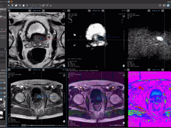
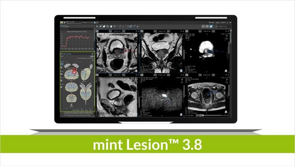
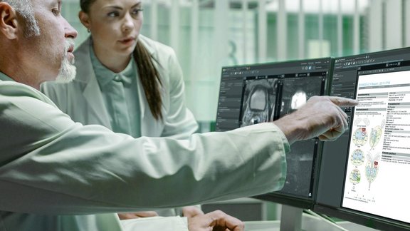
In an often time-consuming multi-parametric prostate MRI read, for instance, mint Lesion™ can automatically import clinical context information into a dedicated and customizable reading profile for the particular job-to-be-done. The clinical data includes PSA values from laboratory reports. These are automatically combined with the volumes of the prostate obtained from the MRI to determine the PSA density. Furthermore, with mint Lesion™ 3.5, measurements of a lesion can be copied to other image series, as the DWI or DCE MRI data. mint Lesion™ also assists in classifying the PI-RADS score of the lesion, and it automatically derives the overall score and index lesion. The resulting report comes in a structured form and includes visualizations and diagrams for reproducibility by referrals. The results can be forwarded as DICOM objects to the urologist and be used as basis for a more precise biopsy. Researchers may correlate the MRI-based results with biopsy information and data from pathological reports by means of mint Analytics. It allows to visually explore interdisciplinary, structured data to achieve advances in the value of image-based diagnostics.
Overall, mint Lesion 3.5 involves AI over the various steps of the radiological read: Beginning with an understanding of the clinical context of the particular patient case to the automatic generation of structured, reproducible reports. Hence, with the help of AI, tomorrow's radiologists will make decisions having their patient's disease data and reports precise and focused. Learn more about mint Lesion 3.5, contact us.


