The desire to standardize reporting and to get even more out of their oncological images led to the partnership of the Noordwest Ziekenhuisgroep in the Netherlands and Mint Medical.
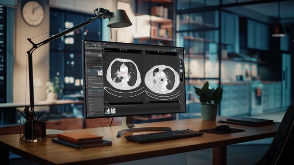
Currently, mint Lesion™ is used in clinical routine in the hospital in Alkmaar: image assessment is done according to RECIST, irRECIST and PI-RADS, with TNM8 being used for colon/rectum. Dr. Remy Geenen, Head of Radiology, is very satisfied with the software solution and convinced of its benefits: “By using mint Lesion™, we are able to generate standardized, complete and consistent oncologic reports. Not only can we describe all the important aspects of a particular tumor, but we are also storing important data in the background, which can be used for quality control and science.” The structured, high-quality annotated data in mint Lesion™ can be used to augment decision-making in multidisciplinary tumor boards or draw up new hypotheses, which go beyond the published literature.
The plan is to gradually expand the use of even more templates in their clinical routine, setting a high standard for their reporting and facilitating interdisciplinary collaboration between colleagues and referring physicians. Since mint Lesion™ generates reports in a clearly structured layout, it is easy for physicians from other disciplines to directly find the information that is relevant to them. This, coupled with the fact that snapshots of findings are integrated directly into the report, leads to fewer queries and makes it possible to communicate better with patients as well.
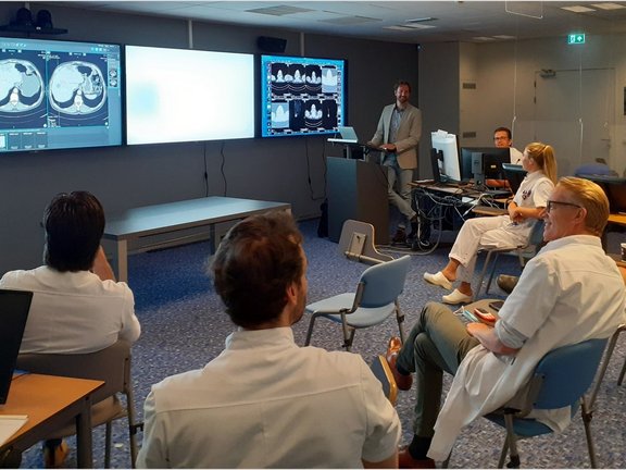
Last month, our colleagues could finally meet up again in person with the radiologists in Alkmaar for a user training session. We look forward to further personal meetings and to our successful partnership!

High-quality annotated data in clinical routine
Related Resources
Related Resources

Lung Cancer Screening in Germany – A Turning Point for Early Detection
Few topics are currently as impactful in radiology as lung cancer screening. From 2026, Germany is expected to roll out a nationwide program, marking…

Standardizing Oncological Imaging: The BORN Project
The BZKF BORN Project (Bavarian Oncological Radiology Network) is setting new standards in the collection and evaluation of oncological imaging data.…

Leave No Data Behind: Commitment to Data Excellence
At Mint Medical, we understand data as a cornerstone for improving patient care and groundbreaking research. Our mission is clear: Leave No Data…