Solid pseudopapillary neoplasms (SPNs), or Frantz tumors, are rare pancreatic tumors accounting for 2-3% of all pancreatic neoplasms. These tumors typically exhibit low malignant potential. Differentiating SPNs from pancreatic neuroendocrine neoplasms (pNENs) using imaging can be challenging due to overlapping features.
Researchers at Heidelberg University Hospital conducted a study to differentiate between SPNs and pNENs using preoperative CT or MRI scans. They analyzed scans of 39 patients with SPNs and 127 patients with pNENs. The mean age of SPN patients was 32.7 years, with a higher prevalence in women, while the mean age of pNEN patients was 51.8 years, with no significant gender difference.
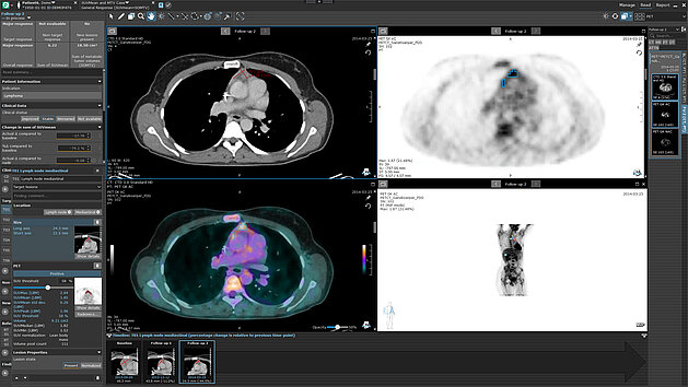
Imaging features such as tumor location, volume, shape, margin, capsule presence, signal intensity/density, and enhancement patterns were assessed using mint Lesion™.
The imaging findings revealed that SPNs typically appeared as round/ovoid lesions with a capsule, more frequently hypointense on unenhanced T1-weighted imaging, and hyperintense on T2-weighted imaging. In contrast, pNENs tended to show lobulated/irregular shapes and arterial hyperenhancement and were associated with upstream pancreatic duct dilation (p < 0.01) and obstructive cholestasis (p = 0.046).
SPNs were more likely to undergo cystic degeneration (p < 0.01) and presented as hypodense on unenhanced CT imaging (p < 0.01). The presence of a capsule was a significant discriminator observed in most SPNs but only a minority of pNENs (p < 0.01).
The study highlights three key features for distinguishing SPN from pNEN: younger patient age, absence of arterial hyperenhancement, and presence of a capsule.
“These discriminating features can be easily applied to routine diagnostics,” state the authors. “They can help to avoid the rather frequent imaging misdiagnosis of these relatively uncommon pancreatic tumor entities which show a significant overlap of epidemiological, clinical, and imaging features.”
Read the original publication here: Imaging differentiation of solid pseudopapillary neoplasms and neuroendocrine neoplasms of the pancreas - ScienceDirect
Khristenko, E, Gaida, MM, Tjaden, C et al. Imaging differentiation of solid pseudopapillary neoplasms and neuroendocrine neoplasms of the pancreas. European Journal of Radiology Open, 2024.
Image above taken from publication.