jump to content jump to footer
Integrated prostate cancer workflow connecting radiology, urology, and pathology through structured reporting, MRI-guided biopsy, longitudinal follow-up, and AI-supported analysis.

Be Central to the Insight: A Structured, Data-Driven Approach to the End-to-End Prostate Pathway

Digital Integration for Multidisciplinary Teams 

In the complex journey of prostate cancer diagnostics, the data contributed by every department is essential to a successful outcome. However, for many clinical teams, the challenge lies in the fragmented nature of data as a patient moves between departments.

mint Lesion acts as your digital infrastructure, allowing you to remain Central to the Insight. The platform connects Radiology, Urology and Pathology by centralizing data and standardizing the reporting process from initial imaging to final clinical diagnosis. This coordination of multidisciplinary insights enables a structured, data-driven experience for the entire clinical team. 

Workflow Integration and FHIR Connectivity

A smooth workflow is defined by how well information flows from one specialist to the next. In many conventional settings, communication bottlenecks can occur because findings are managed in isolation. While urologists often receive static text reports lacking spatial data, pathologists frequently provide results as separate, independent documents. In these siloed environments, correlating histological findings with the original radiological areas or volumes of interest remains a manual process that can lead to fragmented data and a lack of immediate feedback.

mint Lesion provides the digital infrastructure to bridge these gaps, transforming disconnected reports into a unified clinical stream. Radiologists utilize the platform as their central hub, aggregating data to address these barriers and to drive a continuous end-to-end workflow from imaging to diagnosis.

To facilitate this at a technical level, the software supports interoperability with FHIR servers through the exchange of both FHIR Observations and Diagnostic Reports. By adhering to the Implementation Guide developed in collaboration with the Medical Informatics Initiative (MII), mint Lesion promotes high-level data standardization. This connectivity allows the platform to integrate clinical parameters and transfer findings, including PI-RADS scores and volume measurements, directly back to the electronic health record as structured, actionable data.

Please scroll horizontally to view all columns (Phase, Functional Role, Outcome).

Phase Functional Role Outcome
Imaging
(Radiology)
Interpretation of mpMRI scans using PI-RADS v2.1. Radiological Diagnosis (e.g., PI-RADS 4)
Biopsy
(Urology)
Targeted tissue sampling guided by radiological data Imaging-informed biopsy samples with traceable lesion localization
Data Consolidation
(Pathology)
Provision of pathological findings from tissue analysis Imaging-informed biopsy samples with traceable lesion localization
Final Review
(Urology)
Review of all findings and patient history Clinical Diagnosis and Treatment Plan

Standardizing the Radiological Diagnostic Process

The end-to-end approach begins with the standardized assessment of multiparametric MRI (mpMRI). Through the guided interface, radiologists objectively evaluate the peripheral and transition zones based on T2w, DWI/ADC, and DCE sequences, ensuring their assessment follows the PI-RADS v2.1 classification.

mint Lesion integrates with AI software from third-party partners for prostate segmentation, volume calculation, and lesion detection. It imports and displays 3D segmentations of the prostate and lesions, allowing users to review and correct them if necessary. The software also maps each finding onto the standardized prostate lesion scheme, reducing the need for time-consuming manual lesion annotation and drawing.

Furthermore, the integration of the PI-QUAL v2 score allows radiologists to document the diagnostic quality of the scan itself, ensuring that all subsequent decisions are based on high-quality imaging data.

Facilitating the Transition from Radiology to Urology 

One of the most significant bottlenecks in prostate care is the transfer of target locations from the MRI suite to the biopsy room.  

With mint Lesion, radiologists support targeted MRI/US fusion guided biopsy by exporting segmented lesions as DICOM RT-STRUCT or DICOM Segmentation Objects, providing the urologist with exact data, which can be integrated directly into urological biopsy devices. 

This digital hand-off replaces manual sketches or verbal descriptions with precise spatial data, providing urologists with the coordinates identified during your radiological diagnosis and facilitating a closed-loop system that supports diagnostic confidence. 

A lesion contour exported from mint Lesion and displayed within the BiopSee® software
A seamless digital hand-off: A lesion contour exported from mint Lesion and displayed within the BiopSee® software by MedCom for targeted guidance

Closing the Diagnostic Loop: A Unified Clinical View 

mint Lesion supports the integration of radiological, urological, and pathological findings into a unified history, allowing the clinical team to correlate histological results directly with the initial radiological assessments. This coordinated approach provides the multidisciplinary team with a consolidated view of the patient’s diagnostic journey, supporting informed treatment decisions without manual data reconciliation. 

The value of this integrated approach is best seen in its impact on daily clinical collaboration. Dr. Jan Rabe highlights how unifying these disparate data sources enhances the interdisciplinary partnership: 

“I appreciate our collaborative solution because it allows us to map the multi-stage process of prostate diagnostics interdisciplinarily - incorporating laboratory, radiology, urology, and pathology- within a single software platform. The participating departments can access pre-diagnostic data in mint Lesion at any point during the assessment. This facilitates interdisciplinary understanding, streamlines the interpretation of results, and saves us from having to gather data from multiple different sources.” Dr. Jan Rabe, Karlsruhe City Hospital. 

Long-Term Management and Active Surveillance 

As patients enter long-term monitoring, the radiologist’s role transitions from initial detection to longitudinal navigation. mint Lesion enables the tracking of prostate findings over time for MRI-based active surveillance, allowing radiologists to monitor subtle changes and compare historical data with high spatial precision. By providing this evidence-based continuity, you support informed decision-making for patient management and enhance the overall quality of care throughout the surveillance period. 

mint Lesion: end-to-end approach in prostate cancer diagnosis
mint Lesion supports the integration of radiological, urological, and pathological findings into a unified history.

Managing Advanced Disease with Efficiency 

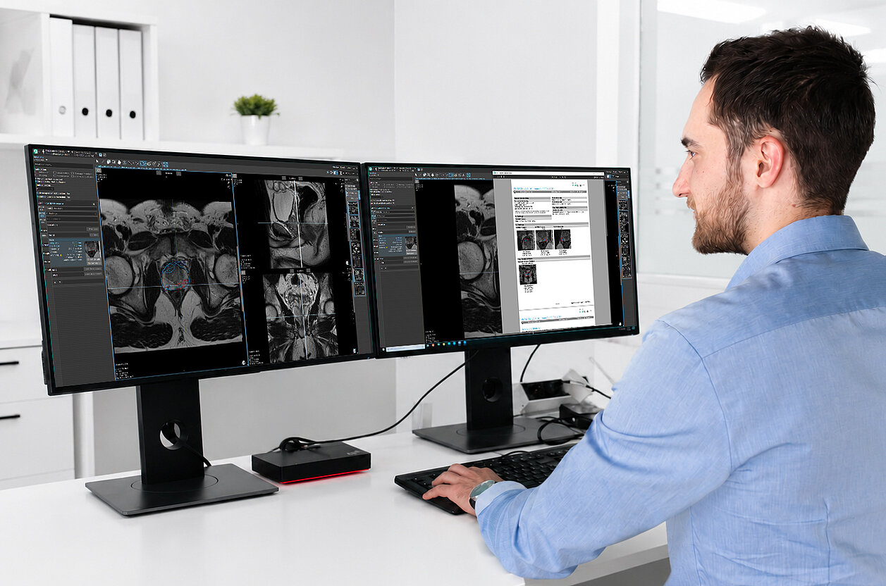
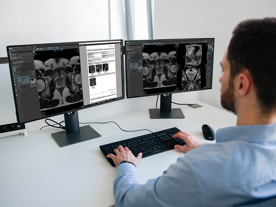
The end-to-end philosophy extends beyond primary diagnosis into the management of advanced prostate cancer. For patients with suspected metastatic spread, mint Lesion provides specialized AI-tools for tumor load quantification in whole-body mpMRI imaging. 

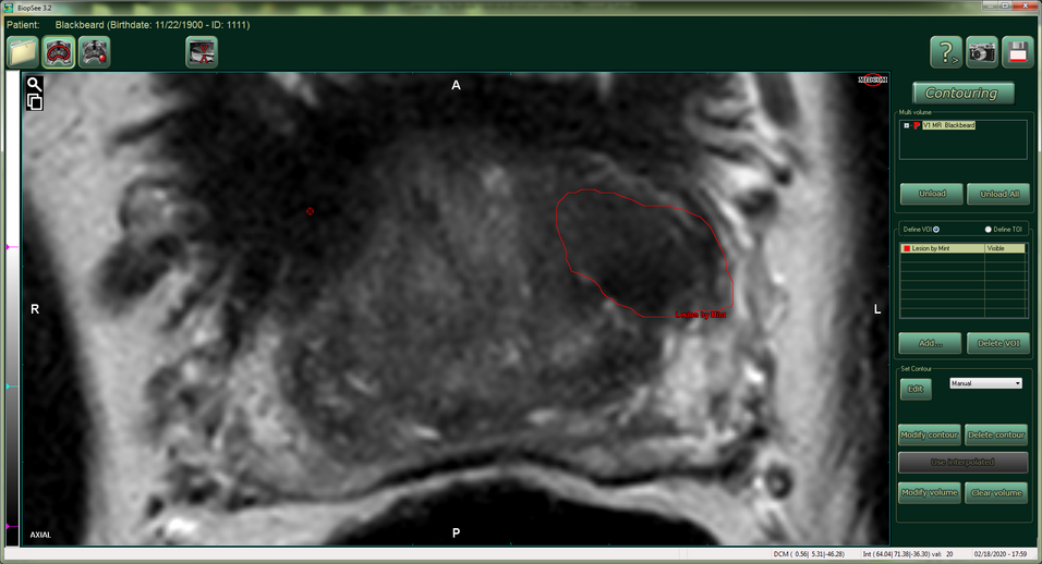
In cases of bone metastasis, manual delineation is a tedious and time-consuming process that often takes an hour or more per scan1, making it impractical for routine clinical use. Consequently, without AI support, assessments remain largely qualitative and inherently less precise.

Even semi-automated tools can require between 12 and 23 minutes1 to finalize results. In contrast, the AI-supported workflows within mint Lesion are designed to execute the entire computation pipeline in approximately 90 seconds1. This significant acceleration allows for the completion of the full automated workflow - including pre- and post-treatment analysis and structured reporting - in approximately three minutes1. This functionality supports the rapid assessment of treatment response, providing the tumor board with structured data to evaluate therapy effectiveness within a standard clinical timeframe. 

By aligning these distinct clinical phases within a single digital environment, mint Lesion moves the diagnostic process beyond fragmented data toward a unified patient history. This integrated approach supports the clinical team in maintaining consistency from the first image to the final treatment plan, ensuring that every diagnostic contribution remains a visible and accessible part of the patient journey. 

Position your expertise at the center of the clinical team with a connected, data-driven workflow. Request a personalized demo to see how you can remain central to the insight. 

Request a personalized demo here.


1Candito, A., Blackledge, M. D., Holbrey, R. Et al. AI-driven software for automated quantification of skeletal metastases and treatment response evaluation using whole-body diffusion-weighted MRI (WB-DWI) in advanced prostate cancer, Phys. Med. Biol. (2025). https://doi.org/10.1088/1361-6560/ae19c5

Radiologist reviewing whole-body MRI images with AI-supported tumor burden quantification for bone metastases and multiple myeloma using mint Lesion.
Mint Medical has integrated an AI algorithm developed by The Institute of Cancer Research and The Royal Marsden into mint Lesion. The solution supports bone disease assessment and treatment response evaluation in advanced prostate cancer and multiple myeloma.
The ICR and The Royal Marsden work with Mint Medical to integrate AI-powered software into cancer treatment
The Institute of Cancer Research (ICR), London, and The Royal Marsden NHS Foundation Trust have announced a new licensing agreement with MedTech…
Read more
Onur Özek from Mint Medical discusses the challenges, requirements, and future of lung cancer screening in Germany in an interview with RadMag.
In the latest RadMag Special Edition, Onur Özek discusses structured reporting, AI integration, second-reader workflows, interoperability, and the importance of end-to-end workflow management for lung cancer.
RadMag Special Edition: Onur Özek on the Challenges of Lung Cancer Screening in Germany
With the nationwide rollout of Germany’s lung cancer screening, radiology providers are facing new challenges. Structured reporting, mandatory second…
Read more
Mint Medical and Coreline teams coming together to announce partnership
Mint Medical and Coreline Soft integrate AVIEW into mint Lesion to support AI-enabled workflows for structured reporting, Lung-RADS assessment, and longitudinal nodule management in lung cancer screening.
Coreline Soft and Mint Medical Announce Partnership to Advance AI-Enabled Lung Cancer Screening
Coreline Soft and Mint Medical today announced a partnership to advance AI-enabled lung cancer screening, following more than six months of technical…
Read more
scroll-top