jump to content jump to footer
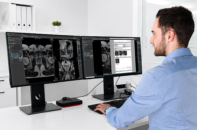
Radiologist analyzing whole-body MRI scans of prostate cancer bone metastases using AI-assisted tumor load quantification in mint Lesion

AI-Supported Tumor Load Quantification for Bone Metastasis in Prostate Cancer

To assess treatment response in patients with advanced prostate cancer, radiologists rely on advanced medical imaging. Conventional modalities, such as bone scans and CT, present limitations in detecting early disease progression and lack standardized criteria for evaluating therapy response [1]. By utilizing Whole-Body MRI, radiologists can detect and monitor metastatic spread patterns, evaluating tumor load non-invasively, radiation free, and without the use of contrast agents or tracers.

However, translating this imaging data into routine clinical insights presents significant workflow hurdles:

  • Manual delineation of bone metastasis is a time-consuming process that often takes an hour or more per scan [2].
  • Even semi-automated tools can require between 12 and 23 minutes to finalize results, making routine use difficult [2].
  • Without structured data, assessing therapy effectiveness across multiple time points can be inconsistent.


AI Capabilities Integrated into mint Lesion

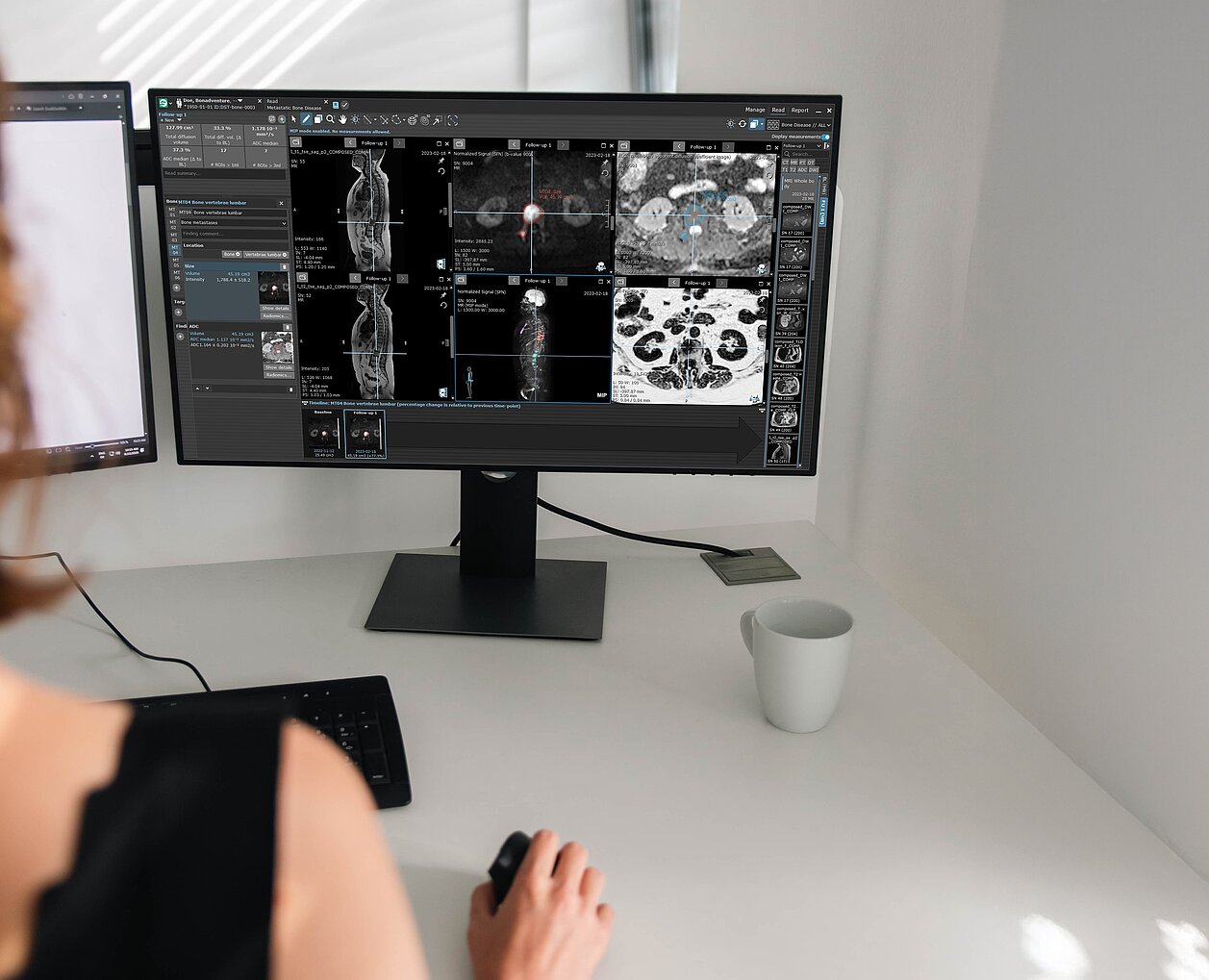
To streamline this process, mint Lesion integrates with a specialized AI algorithm*, developed in collaboration with The Royal Marsden NHS Foundation Trust and The Institute of Cancer Research, London. The algorithm performs the automated image analysis; mint Lesion delivers the quantitative readout, the structured report and the longitudinal comparison.

  • Targeted Data Extraction: mint Lesion presents total diffusion volume (TDV) and global ADC (gADC) statistics for the whole skeleton and for individual body regions in a structured report with color-coded visualizations.
  • Automated Computation: In the published evaluation, the computation pipeline for tumor load quantification completed in approximately 90 seconds [2].
  • Comprehensive Workflow Execution: The published workflow, covering pre- and post-treatment analysis and structured reporting, completed in approximately three minutes [2].
  • Therapy evaluation across time points: mint Lesion compares measurements from multiple time points in a structured framework that supports longitudinal assessment.

mint Lesion implements this workflow within its reading and reporting environment.


Practical Outcomes for Clinical Workflows

Supported by the AI-powered mint Lesion software, radiologists can incorporate full tumor load quantification into standard clinical workflows and maintain reproducible procedures across different scans and time points. Ultimately, this equips them to supply the tumor board with structured, objective data to evaluate therapy effectiveness, providing a factual basis for decisions regarding ongoing patient care.

Interested in AI-supported quantification in other hematologic-oncologic applications? Learn how mint Lesion supports structured assessment in multiple myeloma.


*based on research funded by the National Institute for Health and Care Research (NIHR)


Interested in learning how AI-supported structured reporting can support your clinical workflows? Get in touch with our team to learn more about mint Lesion.


References

[1] Padhani, AR, Lecouvet, FE, Tunariu, N, Koh, DM, De Keyzer, F, Collins, DJ, Sala, E, Schlemmer, HP,  Petralia, G, Vargas, HA, Fanti, S, Tombal HB, de Bono, J (2017). METastasis Reporting and Data System for Prostate Cancer: Practical Guidelines for Acquisition, Interpretation, and Reporting of Whole-body Magnetic Resonance Imaging-based Evaluations of Multiorgan Involvement in Advanced Prostate Cancer. European Urology, 71(1), 81-92. DOI: 10.1016/j.eururo.2016.05.033. PMID 27317091.

[2] Candito A, Blackledge MD, Holbrey R, Porta N, Ribeiro A, Zugni F, D'Erme L, Castagnoli F, Dragan A, Donners R, Messiou C, Tunariu N, Koh DM. AI-driven software for automated quantification of skeletal metastases and treatment response evaluation using whole-body diffusion-weighted MRI (WB-DWI) in advanced prostate cancer. Phys Med Biol. 2025 Nov 14;70(22). DOI: 10.1088/1361-6560/ae19c5. PMID: 41166885.

Mint Medical and Coreline teams coming together to announce partnership
Mint Medical and Coreline Soft integrate AVIEW into mint Lesion to support AI-enabled workflows for structured reporting, Lung-RADS assessment, and longitudinal nodule management in lung cancer screening.
Coreline Soft and Mint Medical Announce Partnership to Advance AI-Enabled Lung Cancer Screening
Coreline Soft and Mint Medical today announced a partnership to advance AI-enabled lung cancer screening, following more than six months of technical…
Read more
Integrated prostate cancer workflow connecting radiology, urology, and pathology through structured reporting, MRI-guided biopsy, longitudinal follow-up, and AI-supported analysis.
mint Lesion supports an end-to-end prostate cancer workflow by connecting radiology, urology, and pathology within a unified digital environment. The platform enables structured mpMRI reporting, MRI/ultrasound fusion biopsy workflows, FHIR-based interoperability, active surveillance, and AI-supported quantification of metastatic disease.
Be Central to the Insight: A Structured, Data-Driven Approach Across the Prostate Pathway
Digital Integration for Multidisciplinary Teams  In the complex journey of prostate cancer diagnostics, the data contributed by every department is…
Read more
Research poster presentation on standardized tumor response assessments using mint Lesion at the NCCN Annual Conference 2026
Presentation of a research poster by Memorial Sloan Kettering Cancer Center on the use of mint Lesion for structured tumor response assessments and clinical research workflows.
NCCN 2026: Memorial Sloan Kettering Cancer Center Presents Research on Structured Tumor Response Assessments with mint Lesion
Big congratulations to Steven Philemond and Alison Chiaramonte of Memorial Sloan Kettering Cancer Center for presenting their research poster “Use of…
Read more
scroll-top