To assess treatment response in patients with advanced prostate cancer, radiologists rely on advanced medical imaging. Conventional modalities, such as bone scans and CT, present limitations in detecting early disease progression and lack standardized criteria for evaluating therapy response [1]. By utilizing Whole-Body MRI, radiologists can detect and monitor metastatic spread patterns, evaluating tumor load non-invasively, radiation free, and without the use of contrast agents or tracers.
However, translating this imaging data into routine clinical insights presents significant workflow hurdles:
- Manual delineation of bone metastasis is a time-consuming process that often takes an hour or more per scan [2].
- Even semi-automated tools can require between 12 and 23 minutes to finalize results, making routine use difficult [2].
- Without structured data, assessing therapy effectiveness across multiple time points can be inconsistent.
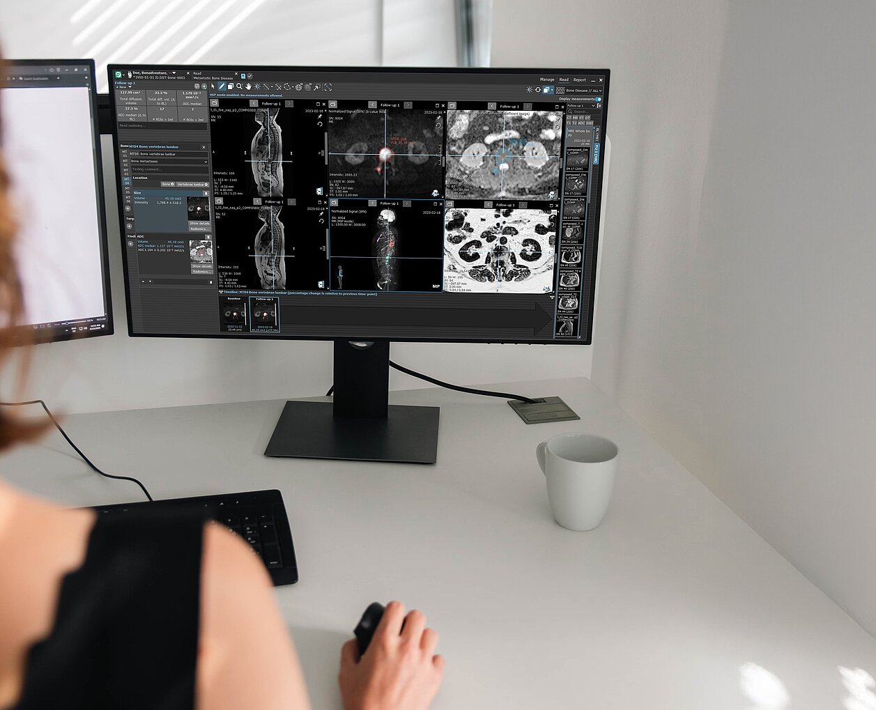
AI Capabilities Integrated into mint Lesion
To streamline this process, a specialized AI algorithm*, developed in collaboration with The Royal Marsden NHS Foundation Trust and The Institute of Cancer Research, London, has been integrated into mint Lesion. This software solution supports the radiologists by addressing core computational and reporting steps:
- Targeted Data Extraction: The software extracts total diffusion volume (TDV) and global ADC (gADC) statistics for both the whole skeleton and specific body regions, presenting these measurements in a structured report with color-coded visualizations.
- Automated Computation: Execute the computation pipeline for tumor load quantification in approximately 90 seconds [2], supported by the AI workflow.
- Comprehensive Workflow Execution: Complete the full automated workflow, including pre- and post-treatment analysis alongside structured reporting, in approximately three minutes [2].
- Consistent Therapy Evaluation: Evaluate treatment response across multiple time points, utilizing a structured framework that facilitates reliable longitudinal insights.
Practical Outcomes for Clinical Workflows
Supported by the AI-powered mint Lesion software, radiologists can incorporate full tumor load quantification into standard clinical workflows and maintain reproducible procedures across different scans and time points. Ultimately, this equips them to supply the tumor board with structured, objective data to evaluate therapy effectiveness, providing a factual basis for decisions regarding ongoing patient care.
Interested in AI-supported quantification in other hematologic-oncologic applications? Learn how mint Lesion supports structured assessment in multiple myeloma.
*based on research funded by the National Institute for Health and Care Research (NIHR)
Interested in learning how AI-supported structured reporting can support your clinical workflows? Get in touch with our team to learn more about mint Lesion.