Multiple myeloma is one of the most complex haematological malignancies – characterized by spatially heterogeneous involvement of the bone marrow that makes reliable diagnosis and monitoring particularly challenging. Conventional methods such as bone marrow trephine biopsy reach their limits here, as they can only ever capture a small sample of the disease.
AI in Whole-Body MRI for Multiple Myeloma
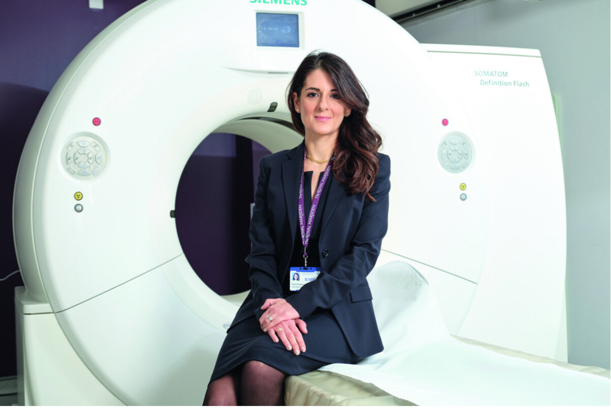
Interview with Prof. Christina Messiou
In recent years, whole-body MRI has established itself as one of the most sensitive and informative imaging techniques for myeloma patients. It provides not only anatomical but also functional and quantitative information – from diffusion-weighted imaging to fat fraction mapping – opening up entirely new possibilities for diagnosis, treatment planning and disease monitoring.
Yet the sheer volume of data generated by whole-body MRI poses significant challenges for radiologists in routine clinical practice. This is precisely where the collaboration between the Institute of Cancer Research, the Royal Marsden and Mint Medical comes in: an AI algorithm integrated into the mint Lesion software automates key evaluation steps – from skeletal segmentation to longitudinal charting – transforming what was once a largely qualitative imaging method into a robust, quantitative tool for precision medicine.
We spoke with Professor Christina Messiou, one of the world's leading experts on whole-body MRI in myeloma patients, about the clinical opportunities as well as the future of this technology.
What are the benefits of Whole-Body MRI for patients with myeloma?
Whole-body MRI (WB-MRI) is a highly sensitive, non-invasive modality for the detection and characterization of myeloma, with demonstrated superiority over low-dose CT and FDG PET/CT for identifying diffuse and focal marrow disease in addition to extramedullary disease. Consequently, the International Myeloma Working Group recommends WB-MRI for surveillance of patients with asymptomatic myeloma and in cases where CT or FDG PET/CT findings are negative or equivocal.
In the UK, WB-MRI is now recommended as a first-line imaging modality for patients with suspected myeloma, supported by both clinical outcome data and health economic analyses. Emerging evidence also supports its routine use in post-therapy assessment, as the presence of residual disease on WB-MRI is associated with inferior outcomes.
The strength of WB-MRI lies in its ability to combine anatomical imaging with quantitative functional techniques, including diffusion-weighted imaging (apparent diffusion coefficient) and Dixon-based relative fat quantification. However, in routine clinical practice, comprehensive extraction and utilization of these quantitative metrics remain challenging.
The collaboration involving The Institute of Cancer Research, The Royal Marsden and Mint Medical integrated an AI algorithm for WB-MRI into mint Lesion. From a practical perspective, how does translating this algorithm into a structured software framework affect the standard reading process for evaluating bone involvement in multiple myeloma?
The integration of AI-driven analysis within a structured reporting platform represents a significant advancement in WB-MRI interpretation. For radiologists, structured frameworks provide consistency and act as a cognitive aid, ensuring comprehensive and standardized reporting of clinically relevant findings.
For hematologists, structured reports improve clarity and accessibility, facilitating efficient navigation and interpretation. The inclusion of visual summaries and infographics enhances multidisciplinary communication and supports patient engagement by improving understanding of their own disease.
Importantly, this approach also enables the systematic capture of high-quality, standardized data, transforming routine clinical imaging into a scalable and mineable dataset for research, biomarker development, and future discovery
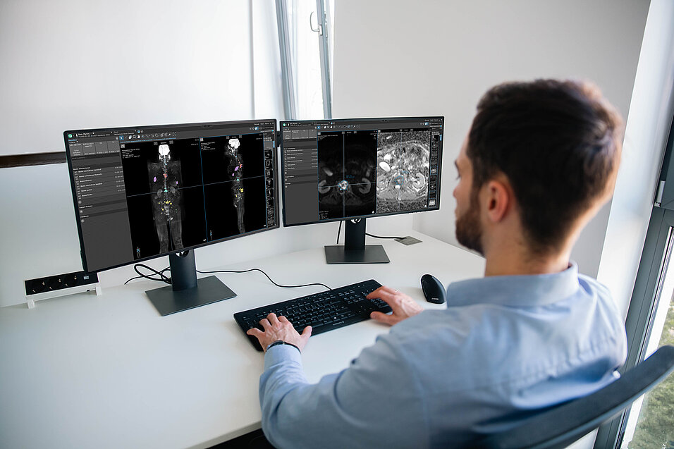
To address the workflow challenges, mint Lesion incorporates automation and AI tools to segment the skeleton into distinct regions, calculate total disease volume, map quantitative metrics like ADC and fat fractions, and align serial scans side by side. From a practical standpoint, how do these automated capabilities change the way a radiologist interacts with a complex whole-body MRI dataset?
Manual segmentation of the skeleton and extraction of regional or whole-body disease metrics are impractical in routine clinical workflows, limiting the full clinical utility of WB-MRI. Automation addresses this barrier by performing these labor-intensive tasks efficiently and reproducibly.
By segmenting the skeleton into anatomical regions, quantifying total disease burden, mapping quantitative parameters such as ADC and fat fraction, and enabling side-by-side comparison of serial studies, AI-driven platforms streamline image interpretation.
This shifts the radiologist’s role from data extraction to data interpretation, providing a structured and intuitive environment in which complex datasets are pre-processed and clinically actionable insights are readily available. In doing so, WB-MRI evolves from a predominantly qualitative tool to a robust, quantitative imaging biomarker platform.
Why do these specific automated metrics, such as regional disease burden tracking, per-region cellularity values, and objective longitudinal chart, matter so much clinically when managing a patient's treatment path over time?
Myeloma is characterized by spatially heterogeneous bone marrow involvement, making sampling with bone marrow trephine inherently limited and prone to sampling error. WB-MRI provides a comprehensive assessment of disease burden across the entire skeleton, and automated quantification enables objective and reproducible measurement of this burden.
As therapeutic advances have extended survival, myeloma is increasingly managed as a chronic condition, often requiring multiple lines of therapy. This necessitates nuanced, data-driven decision-making that balances treatment efficacy with toxicity and patient comorbidities.
Quantitative metrics—such as regional disease burden, cellularity estimates, and longitudinal trends—can provide objective measures of response and progression. By presenting these data in an accessible and standardized format, automated platforms reduce subjectivity and support more precise, personalized treatment strategies over time
As multiple myeloma management increasingly shifts toward long-term disease control, what is your vision for the future of AI-assisted imaging? Specifically, how must software frameworks evolve to integrate emerging quantitative biomarkers and further support precision medicine?
The future of myeloma imaging lies in the integration of multimodal, quantitative biomarkers to better characterize complex disease biology. Single-parameter assessments are increasingly insufficient; instead, comprehensive diagnostic models incorporating multiple imaging and clinical biomarkers will be required.
AI-enabled platforms will play a central role in this transition by facilitating the shift from qualitative to quantitative imaging and enabling scalable integration of diverse data types. Beyond conventional imaging metrics, there is significant potential to incorporate emerging biomarkers, including body composition analysis and predictive imaging signatures.
To realize this vision, software frameworks must evolve to support interoperability, standardization, and seamless integration into clinical workflows. Ultimately, these advances will underpin precision medicine approaches, enabling more accurate risk stratification, treatment selection, and longitudinal disease monitoring in patients with myeloma.
Our conversation with Professor Messiou makes clear just how much imaging in multiple myeloma is currently evolving: away from purely qualitative assessment and towards a data-driven, quantitative tool that enables physicians to make better-informed decisions throughout the entire treatment pathway. As myeloma is increasingly understood as a chronic condition requiring multiple lines of therapy, objective and reproducible longitudinal data are becoming ever more important – both for patients and for the clinical teams treating them.
We would like to sincerely thank Professor Christina Messiou for her time and for these fascinating insights into her clinical and scientific work.
Professor Christina Messiou is a Consultant Radiologist at The Royal Marsden NHS Foundation Trust and Professor at the Institute of Cancer Research, London, where she has worked since 2007. As an oncology radiologist specializing in imaging of myeloma, melanoma and soft tissue sarcoma, she is internationally established as a leading authority on whole-body diffusion-weighted MRI and was among the first radiologists to introduce this technique into routine clinical practice for myeloma patients. In recognition of her research, she was awarded a Roentgen Professorship by the Royal College of Radiologists in 2014 and Royal College of Radiologists and NIHR Clinical Research Network Outstanding Clinical Radiology Researcher Award in 2019. She currently leads the Imaging and Data Science theme of the NIHR Biomedical Research Centre at The Royal Marsden and The Institute of Cancer Research, and was conferred with the title of Professor in October 2022.
Learn more about AI-supported quantification of bone involvement in multiple myeloma with mint Lesion.
Related Resources
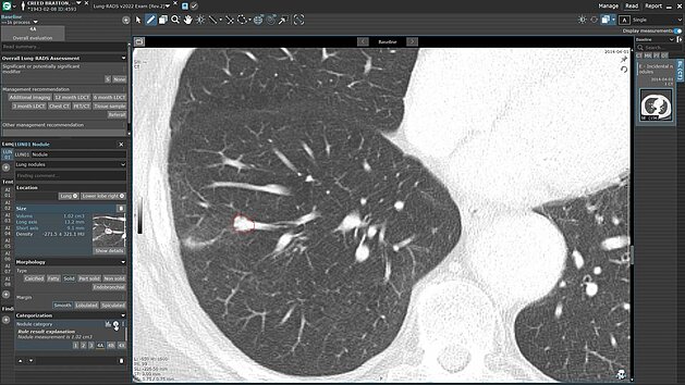
Leverage Advanced AI-Driven Nodule Detection and Analysis for Comprehensive Patient Care
Discover the power of streamlined AI-driven lung screening with contextflow ADVANCE Chest CT integrated into mint Lesion. With automated lung nodule…
Read more

Comparison of iRECIST and RECIST 1.1 for Evaluating Immunotherapy in Melanoma and Non-Small Cell Lung Cancer
In a retrospective study conducted at the University Hospital Cologne, the radiological criteria iRECIST and RECIST 1.1 were compared for assessing…
Read more

Developing Standardized Reporting Templates for Sarcomas: Insights from Prof. Dr. Wolfgang Kunz
Sarcomas are a rare and heterogeneous group of malignant tumors that pose significant challenges in both diagnosis and treatment. Within the framework…
Read more