A retrospective study [1] conducted by researchers at University of Miami Miller School of Medicine and Nova Southeastern University investigated whether quantifying the heterogeneity of the MR signal intensity using image texture analysis and T2-mapping could provide more objective imaging biomarkers for response monitoring in superficial fibromatosis.
Superficial fibromatosis presents a particular challenge in objectively assessing the efficacy of treatment because changes in nodule composition can occur independently of changes in size. Moreover, nodules in this disease are often small and, consequently, absolute changes of only a few millimeters result in large percentage differences, making meaningful longitudinal comparisons difficult.
“Decreased T2 signal in the nodule during the course of therapy likely reflects a transition from the proliferative to a more quiescent phase marked by increased collagen deposition, and may be a better indicator of treatment response than decrease in size,“ explained the authors. “[…] to our knowledge, this is the first study to show that T2 mapping and radiomics analysis can detect longitudinal differences corresponding to biologic maturation in superficial fibromatosis.”
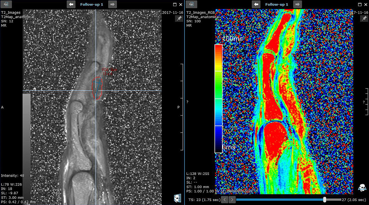
Baseline and follow-up MRI from 13 patients were evaluated. mint Lesion™ was used to draw and analyze regions of interest (ROI) in the regionally largest or most conspicuous nodule on proton-density MRI and corresponding T2-maps, when available. ROIs were also drawn in nearby muscle tissue and served as an internal reference for normalization and comparability of nodal signal intensity.
Comparisons were made between baseline and follow-up T2 relaxation times and radiomics features: entropy, kurtosis, skewness, mean of positive pixels (MPP), and uniformity of distribution of positive gray-level pixel values (UPP), which were calculated from the ROIs using the mint Lesion™ specific settings, implemented in accordance with the “Image Biomarker Standardization Initiative”.
Normalized T2 signal intensity and T2 relaxation time showed significant decrease (23%, p = 0.03, and 16%, p < 0.001), while skewness increased (73%, p = 0.03) and entropy decreased (4%, p = 0.05).
“Our findings support use of these measurements as novel imaging biomarkers of activity in superficial fibromatosis, and hint at the possibility of broader relevance in other fibroproliferative diseases,” concluded the researchers.
[1] Ramachandran A., Fox T., Wolfson A., Banks J., Subhawong T.K. (2021) Superficial fibromatosis: MRI radiomics and T2 mapping correlate with treatment response. Magnetic Resonance Imaging. doi.org/10.1016/j.mri.2021.06.003