An exploratory study [1] conducted by researchers at University Hospital Tuebingen investigated whether CT texture analysis parameters correlate with semiquantitative 18F-FDG PET parameters and therapy relevant mutations of tumor cells in metastatic melanoma, assessed by the histopathologic specimen.
Overall survival of patients with advanced melanoma is still limited and could be significantly improved by targeted therapies. However, the therapeutic outcome depends on the mutational status, and the individual treatments rely on genomic analysis of tumor cells – which requires biopsy and time-consuming histopathological assessment. So, “any imaging technique providing information on mutational status will be a tremendous advantage in melanoma, noted for its rapid rate of spread,” explained Prof. Dr. Klumpp.
Currently, 18F-FDG PET/CT provides the most accurate results for staging, follow-up, and pre-surgical evaluation of patients with advanced melanoma. Still, CT texture analysis has proven to be useful for tissue characterization, for prediction of mutational status, and of patients’ prognosis in oncological imaging in several tumor entities.
“This retrospective study attempted to correlate CT parameters with mutational features of metastatic melanoma on histopathology,” the authors declared.
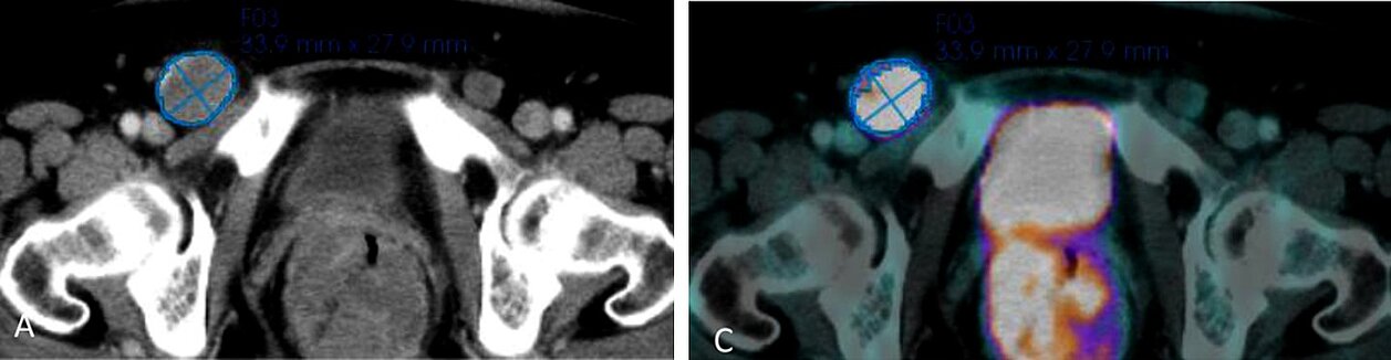
Data sets from 66 patients with one metastasis (53 patients) or several metastases (13 patients) were evaluated. mint Lesion™ was used to assess the largest metastasis in each patient and to extract:
- CT texture parameters (density, attenuation standard deviation (SD), kurtosis, skewness, mean value of positive pixels (MPP), uniformity of distribution of positive pixels (UPP), entropy, and uniformity) and
- Semiquantitative 18F-FDG PET parameters (volume and mean and peak values of the standard uptake value (SUV)).
Tissue samples were analyzed for the B-RAF gene, NRAS gene, and wildtype.
A negative correlation was obtained for attenuation SD with SUVpeak (rho -0.292, p 0.017), and no relationships between the other CT texture analysis parameters and semiquantitative 18F-FDG PET parameters were found.
Attenuation SD correlated with SUVmean in BRAF- (rho 0.432, p 0.050) and NRAS-mutation group (rho -0.742, p 0.008). It also correlated with SUVpeak in NRAS-mutation (rho -0.790, p 0.003) and wildtype-patients (rho -0.427, p 0.033).
“CT texture parameters cannot replace the diagnostic value of 18F- FDG PET/CT for metabolic information in melanoma patients,” concluded the researchers. “Large scale prospective trials with highly standardized inclusion criteria, scanner types, and protocols might help to identify potentially clinically relevant associations of CT texture analysis parameters with tumor characteristics.”
[1] Olthof S. C., Krumm, P, Weichold, O., Eigentler, T., Bösmüller, H., la Fougère, C., Pfannenberg, C., Martus, P., Klumpp, B. CT texture analysis compared to Positron Emission Tomography (PET) and mutational status in resected melanoma metastases, European journal of Radiology, Vol. 131, Oct 2020, https://doi.org/10.1016/j.ejrad.2020.109242