We all have been exposed to the riveting news of artificial intelligence tools taking over diagnostic imaging. Due to the rapid increase in global drivers such as aging population, new government initiatives and growing imaging technology, there is a pressing need to address the increased general radiologist’s workload as well as the efficiency and quality of the expert readers. The number of imaging specialists is simply not keeping up with the increasing demands of diagnostic workups.
At Mint Medical, we understand that artificial intelligence tools will play a critical part in the daily work of a radiologist. In a role of an intelligent assistant, deep learning and knowledge modeling tools will increase diagnostic read efficiency by helping physicians automate and standardize their mundane tasks and free up the time for complex diagnosis.
“At Mint Medical, we understand data. We were one of the first companies to develop structured guided reports for oncology tumor response assessment. Today, we continue pioneering the area of intelligent diagnostic assistance through deep learning techniques within and beyond the oncology field,” says Dr. Matthias Baumhauer, company’s founder and CEO.
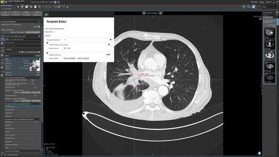
At the 104th annual meeting of RSNA in Chicago this year, mint Lesion will debut its artificial intelligence assistant prototype that utilizes a multitude of AI techniques (deep-learning technique based on a modified, U-Net Convolutional Neural Network, trained on hundreds of thousands of ground-truth data, is just one of them).
Let’s take for example a lung-cancer patient with metastatic disease who has undergone treatment for the past five years at different facilities. All of the reports are unstructured with no clear indication of lesion slice location or standardized lesion measurements, making it almost impossible to track this patient’s progress accurately and deterministically. With mint Lesion 3.5 prototype, radiologist can rely on mint Lesion AI assistant to automatically measure prior studies and to have prepared critical components of the final report content (in a single and multi-modality setting). The software will, subsequently, take the one-dimensional measurements and turn them into volumetric calculations of the corresponding prior lesions. Instead of taking five-fold the time to identify all of the prior lesions and decipher non-standardized prior measurements, the reader will have all the longitudinal workup prepared by mint Lesion.
With this standardized approach, the outcome of the reader’s report will have clearly communicated, objective and reproducible content that referring physicians can draw from, whether in a clinical trial scenario, clinical routine or research.
Quality of patient’s outcomes is likely to be improved dramatically as well as it should be much easier to confirm that the selected reference lesions represent disease, to affirm response to treatment or better identify more comprehensive responses to treatment or ambiguous response situations.
Each facility that decides to utilize such a tool prospectively in their clinical routine will have a chance of conducting deep learning work and prognostic studies’ research in a multitude of ways. As one may predict, down the road, this offers room for real-time AI assistant and potential identification of mis-diagnosis (i.e. reduction of false negatives).