jump to content jump to footer
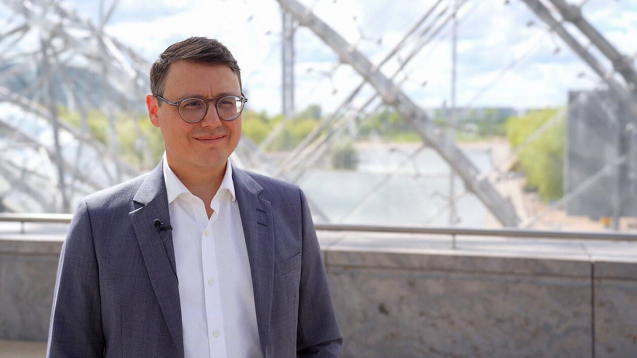
Portrait of Prof. Dr. Timm Denecke, Director of Radiology at Leipzig University Hospital, RÖKO 2026

RACOON-MARDER: AI Research for the Early Detection of Hepatocellular Carcinoma

Interview with Prof. Dr. Timm Denecke

Hepatocellular carcinoma (HCC, liver cancer) is one of the most common malignant tumors worldwide. Early detection and precise assessment of disease progression are crucial for selecting the appropriate treatment and achieving successful outcomes. We know which liver diseases are associated with an increased risk of developing HCC, and there are corresponding, relatively pragmatic recommendations for HCC screening, which is primarily based on ultrasound. But how can patients at risk be stratified more precisely in the future and, in cases of high risk, referred for more intensive early detection? And how can artificial intelligence and structured imaging data contribute to a more precise assessment of an individual’s HCC risk?

At RÖKO 2026, we had the opportunity to speak with Prof. Dr. Timm Denecke, Director of the Department of Diagnostic and Interventional Radiology at Leipzig University Hospital, about his research. Leipzig University Hospital is a liver transplant center with a strong focus on liver diagnostics and hepatology.

As a recognized expert in liver radiology, Prof. Denecke is actively involved in the Network University Medicine (NUM) and, in particular, the radiological research network RACOON (Radiology Research Network). In this interview, he provides insights into his research project RACOON-MARDER, explains its objectives, and discusses how structured imaging data and artificial intelligence can contribute to research into hepatocellular carcinoma.

Prof. Denecke, what is behind the name RACOON-MARDER?

The project I came up with is called RACOON-MARDER.

MARDER is an acronym that essentially stands for AI research using liver MRI data. Like its parent project RACOON, the name draws inspiration from the animal kingdom. That is how it came about.

With MARDER, we want to leverage the Network University Medicine and RACOON to rapidly compile large volumes of carefully selected, standardized data that consistently represent the same scenario: patients at increased risk of developing hepatocellular carcinoma (HCC). This increased risk is generally associated with chronic liver disease, such as liver cirrhosis or liver fibrosis.

Our aim is to capture the stage before a malignant tumor develops and investigate whether artificial intelligence can be used to predict HCC. In other words, we want to assign an individual risk to a patient in advance of developing hepatocellular carcinoma later on.

Why is this so important? Patients at increased risk are included in screening programs, and this involves a very large number of people. As a result, such screening is resource-intensive and costly when only a comparatively small number of tumors are ultimately detected.

At present, screening is generally performed using ultrasound, including without ultrasound contrast agents or similar methods. The challenge, however, is that it is not always possible to assess the entire liver or to detect small tumors at an early stage, when they would be particularly amenable to treatment.

Of course, these patients could instead be examined using a more advanced imaging modality such as MRI. While MRI does not involve ionizing radiation, it is considerably more expensive. This means investing substantial resources to identify a relatively small number of cases in which hepatocellular carcinoma can be treated earlier and more effectively.

There is also a second challenge: MRI is a limited resource. Both the scanners themselves and the professionals who operate them—our medical technologists—are in short supply. We simply would not have the capacity required to provide MRI examinations on this scale.

The key, therefore, is to identify a subpopulation whose risk of developing hepatocellular carcinoma is high enough to justify regular examinations using MRI, despite its cost and limited availability. This is precisely where our work begins. We aim to predict this individual risk by using AI to analyze existing MRI images.

What role does artificial intelligence play in the project?

I don't think this project would work without AI.

What we are looking for are likely precursor lesions of HCC. These can be detected in certain contrast-enhanced MRI examinations and are known as dysplastic nodules.

However, the progression from a dysplastic nodule to liver cancer can sometimes occur much more rapidly or may even skip certain intermediate stages. We can only hypothesize about this because we do not detect such a nodule beforehand in every case.

It is also important to understand that a cirrhotic liver is full of nodules of all kinds of sizes, shapes, colors, or shades of gray—after all, much of what we see in radiology is black and white. Amid all this noise, the crucial nodule may simply escape the human eye.

This is precisely where we are relying on artificial intelligence. With its enormous computational power, AI is intended to find the proverbial needle in the haystack and identify findings that indicate a lesion may be progressing toward malignant transformation into hepatocellular carcinoma—earlier than would be possible for the human eye.

Video interview Dr. Timm Denecke, RACOON-MARDER
Watch Prof. Dr. Timm Denecke talk about how RACOON-MARDER uses AI-analyzed MRI to predict hepatocellular carcinoma on Youtube (English subtitles available).

What challenges need to be overcome in RACOON-MARDER?

In my opinion, the greatest challenge is not necessarily the subsequent development of the algorithm itself, but first assembling the training cohort—the population on which the algorithm will be trained.

For this, we need a large number of carefully selected MRI datasets. All of the scans must demonstrably have been acquired before the development of hepatocellular carcinoma. At the same time, these patients must subsequently have been diagnosed with HCC during follow-up.

However, that is only the test cohort. We also need a control group in which no hepatocellular carcinoma developed within a defined period, for example, three to five years.

Overall, we have calculated that we will need approximately 600 to 800 cases, roughly half from the control group and half from the positive cohort, in order to train the algorithm meaningfully.

That is a major challenge. This is why I am relying on the strength of the network—or, if you like, on a kind of “crowdfunding” of cases. That is exactly what we depend on for this project.

How do the technical partners within the network support the project?

The network supports us primarily by connecting virtually all university medical centers in Germany. Only a network of this scale makes it possible to compile a sufficient number of carefully selected cases.

In addition, there are technical partners that enable both the operation of the network and the secure exchange of imaging data via the respective nodes. Mint Medical, for example, is one of these partners. For the project-specific research questions (in particular, the primary and secondary endpoints of our study) we are also supported by MEVIS.

What milestones has the project already achieved?

We are still at the beginning of the project. However, reaching this point has already required a great deal of preparatory work.

One important milestone was obtaining ethics approval, which has now been granted. It can now be applied throughout the network for all participating partners, in principle, all university hospitals in Germany. This was a crucial hurdle that we have already overcome.

Having also established the technical infrastructure and defined and allocated the work packages, we are now heading directly toward the kick-off. This means that we will soon be able to begin data collection.

What motivates you, and what do you hope for the future?

What excites me about the project is, of course, first and foremost the scientific objective we hope to achieve. This also includes the scientific output one hopes for as a researcher, as well as the opportunity for the many people involved in the project to benefit from it personally and scientifically.

But above all, our patients are at the heart of this project. We want to help them through the project and the algorithm that will ultimately emerge from it.

What particularly excites me beyond that is something I have never experienced on this scale during my scientific career: this highly integrative collaboration.

In keeping with the theme of this year's German Radiology Congress, it really is almost without boundaries. People support one another without restrictions, exchange ideas openly, and work together toward the same goals.

Large network projects like this simply would not be possible otherwise. After all, MARDER is only one of many projects currently being funded within RACOON. We depend on one another.
Our conversation with Prof. Dr. Timm Denecke highlights the potential of structured imaging data, artificial intelligence, and interdisciplinary research networks to advance liver diagnostics. We would like to sincerely thank Prof. Denecke for these fascinating insights into the RACOON-MARDER research project and wish the entire project team continued success.

RACOON-MARDER is one of seven projects within Topic Area 6 of NUM 3.0. In addition to MARDER, BRAIN-AI, COMPARE, INCLUDED, LCS, PAIN, and PROSTAIT are also receiving funding. The project is funded by the Federal Ministry of Research, Technology and Space (BMFTR) within the Network University Medicine (NUM) as a use case of the Radiological Cooperative Network (RACOON).

The project brings together a broad range of partners. In addition to Leipzig University Hospital, the core team includes Hannover Medical School, represented by PD Dr. med. Anna Saborowski and Prof. Dr.-Ing. Andrea Schenk; Prof. Dr. med. Dominik Geisel from Charité Berlin; and LMU Munich, under the leadership of Prof. Dr. med. Sophia Stöcklein. On the technology side, the project is supported by Fraunhofer MEVIS, the German Cancer Research Center (DKFZ), and Mint Medical.

To learn more about the ongoing projects, visit the Network University Medicine (NUM) website or the RACOON Network website.

Or explore more interviews about RACOON projects here.

RACOON FADEN Project Tackles Early Detection of Adenomyosis
Endometriosis is a vastly under-researched condition affecting women, but it is finally receiving the attention it deserves through the RACOON FADEN…
Read more
Early Detection of Treatment Response in Lung Cancer Using Delta-Radiomics Features
The study conducted by Heidelberg University Hospital investigates the use of diffusion-weighted MRI (DWI) to predict early treatment outcomes in…
Read more
scroll-top