In neuro-oncology, imaging assessments directly shape treatment decisions. For example, under RANO 2.0, whether a follow-up MRI is classified as progression can determine if a patient continues on therapy or moves to a different line of treatment. The reliability of that decision depends on consistency: across readers, across time points, and across evolving response criteria. mint Lesion is designed to make glioma MRI reporting structured and reproducible, so referring clinicians can rely on it from first detection through long-term follow-up and on to the tumor board.
The challenge: inconsistency in glioma assessment
Published data points to several sources of inconsistency in glioma MRI assessment. Reader variability of around 22% has been reported for measurements of the same tumor, and readers agree poorly on segmentation after resection¹. Two-dimensional measurements are associated with a false-positive progression rate of around 14%².
Free-text reports add a further gap, as they do not reliably capture every diagnostic item a complete assessment requires. Because RANO 2.0 progression rules drive treatment changes, applying them correctly and consistently is essential.
The Response: consistency by design
mint Lesion addresses these challenges with an approach built on structured reporting, embedded response criteria and reusable data.
Structured reporting and longitudinal tracking
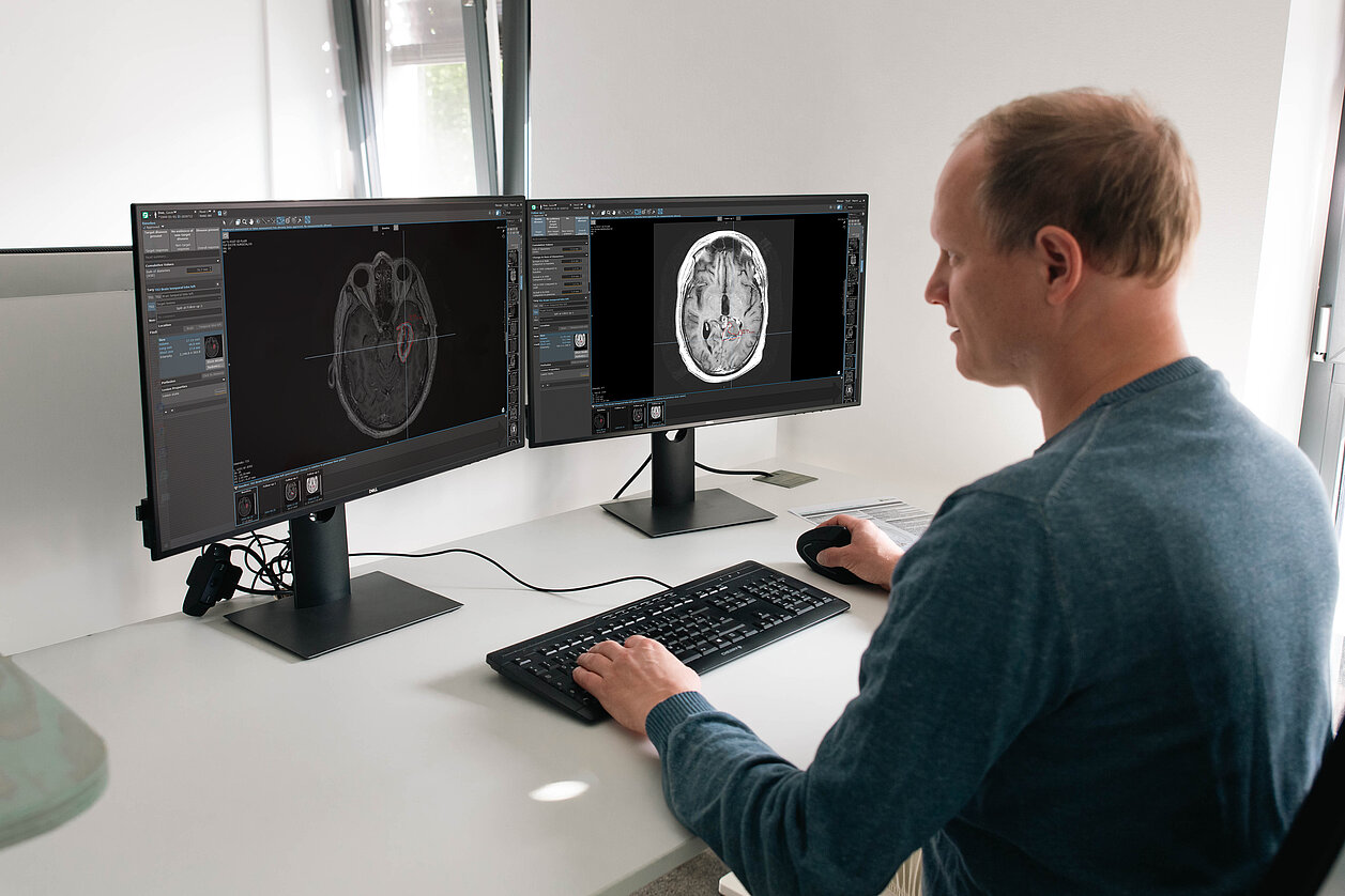
Standardized descriptors and a structured workflow support reproducible assessment. Each measured lesion is followed across examinations, which keeps evaluations consistent from baseline through every follow-up.
Embedded RANO 2.0 and response automation
mint Lesion automates response classification and applies criteria consistently. Parameters can be configured to match the study protocol. Tumor burden is calculated as SPPD (2D) and SOV (volumetric), grouped by lesion type and measurement method. Confirmation-scan logic runs automatically, using RANO 2.0 defaults or custom study settings.
For a closer look at the configuration options, tumor burden tracking and confirmation-scan logic, read our article Implementing RANO 2.0 for Neuro-Oncology Clinical Trials in mint Lesion.
Structured data for downstream use
The data export combines radiomics, annotated images and guideline context. It runs automatically and is ready for statistical analysis, while configurable eCRFs capture non-imaging parameters. Annotated datasets stay linked over time. Supporting DICOM-SEG export and FHIR interoperability, mint Lesion can integrate with radiotherapy planning and the wider clinical IT environment.
Looking ahead: anatomical context and AI-based segmentation
Further capabilities are currently in development*. The Anatomical Patient Model (APM) provides anatomy segmentation, anatomical landmark localization and tumor segmentation for the brain. Its “Anatomy GPS” displays anatomical labels wherever the user clicks and links questionnaires to images, so reports remain traceable.
Building on the APM, AI-based segmentation of brain tumors will provide both the tumor core on T1-weighted MRI and the whole tumor on T2/FLAIR. The aim is to reduce variability between readers and to deliver standardized tumor segmentations as the basis for 2D and volumetric measurements, together with the anatomical context of each tumor.
Reproducible glioma reporting
With structured reporting, embedded RANO 2.0 logic and data ready for downstream use, mint Lesion supports a consistent standard across readers, time points and changing criteria.
* Features in development are not yet commercially available.
References
¹van der Loo, I., Tareco Bucho, T. M., Hanley, J. A. et al. Measurement variability of radiologists when measuring brain tumors, Eur. J. Radiol. (2025). https://doi.org/10.1016/j.ejrad.2024.111874
²Provenzale, J. M., Ison, C., DeLong, D. Bidimensional measurements in brain tumors: assessment of interobserver variability, Am. J. Roentgenol. (2009). https://doi.org/10.2214/AJR.09.2615