Modern technology advancements, specifically in the area of medical imaging, have substantially improved the evaluation of therapy response and the ability to monitor disease status post-treatment in childhood cancer. Yet there remain challenges that create barriers to the overall benefit of childhood cancer research advances.
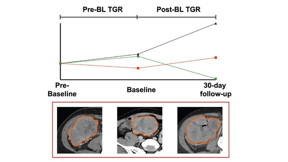
For example, discrepancies in the modalities employed and evaluation frequency, inconsistencies in techniques and assessment protocols, as well as data siloing hinder high-quality evidence-driven analysis in clinical trials. More specifically, diameter-based evaluations continue to be utilized to quantify treatment response and inform treatment efficacy. But such measurements have been shown to demonstrate variability, and with merely this level of quantitation, valuable information that could actually be investigated deeper is often left behind or unacknowledged. There is clearly a need for more consistency and standardization when it comes to collecting and analyzing the vast amount of data provided by medical images. In addition, using other means, such as volumetric measurements, tumor growth kinetics, and radiomic features might be a better way to quantify treatment response and evaluate treatment efficacy. And that is where our software platform, mint Lesion™ comes to play.
As part of our commitment to accelerating progress against childhood cancer, we continue developing a variety of dedicated radiology and clinical data assessment templates that support the evaluation of conventional therapies, immunotherapies, and novel treatment mechanisms for different childhood cancer types, such as High and Low Grade Gliomas, Diffuse Intrinsic Pontine Glioma, Sarcoma, Melanoma, Neuroblastoma, Lymphoma and more. While our mint Lesion™ templates support the application of traditional diameter-based 2D measurements specified by the particular response assessment criteria, at the same time, they allow volumetric measurements, tumor growth modeling, and concurrent extraction of radiomics during any evaluation. Moreover, advanced import/export of DICOM annotations (e.g., NRRD, DICOM SR, DICOM segmentation objects) supports full data liquidity for AI and machine learning. By capturing and integrating all information into a digital model of the patient and its evolution over time, the "minted" radiology report provides a meaningful, comprehensive artifact of the image evaluation results for the physician provider and the patient.
These and other mint Lesion™ features ensure no data is left behind on the image and provide clinicians with more detailed information to guide childhood cancer research and validate the optimal evaluation measures.
To support the advancement of childhood cancer research, we at Mint Medical focus on providing the path to structured, mineable, semantic data, creating a solid basis for evidence-based research. We believe that combining therapy response assessment with the framework for a more profound investigation of the imaging features significantly increases the prospects of identifying better avenues for childhood cancer treatment. With our technology, we remedy data siloing and loss of context to information as it makes its way from the patient to the image to the provider and other stakeholders in the life cycle of the results abstracted from the image.