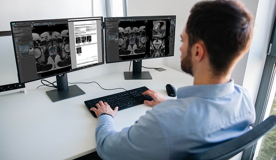
mint Lesion: oncology imaging software for guided reading, structured reporting, longitudinal data and multi-center research
mint Lesion is a radiology software platform from Mint Medical that combines image viewing, guided assessment based on established criteria and classification systems, and automated structured reporting. Radiologists use it to follow disease status and treatment response over time, imaging researchers to run single- and multi-center biomarker studies from protocol design to AI-ready datasets, and trial sites, pharma companies, and CROs to capture standardized, longitudinal data for clinical trials.
Where is mint Lesion used?
One platform for different settings, each with its own reading workflows and data requirements.

Diagnostic radiology
Guided reads and automated structured reports for screening, staging, and follow-up.

Clinical trials
A reading platform for consistent, audit-ready therapy response assessment at trial sites.

Multi-center research
Consistent protocols and standardized data across all participating centers, ready for pooled analysis and AI.

Pharma and CROs
Reproducible imaging endpoints across sites, readers, and time points.
What does mint Lesion do?
mint Lesion guides readers through each assessment, keeps every finding connected over time, and turns the read into a report and a reusable dataset.
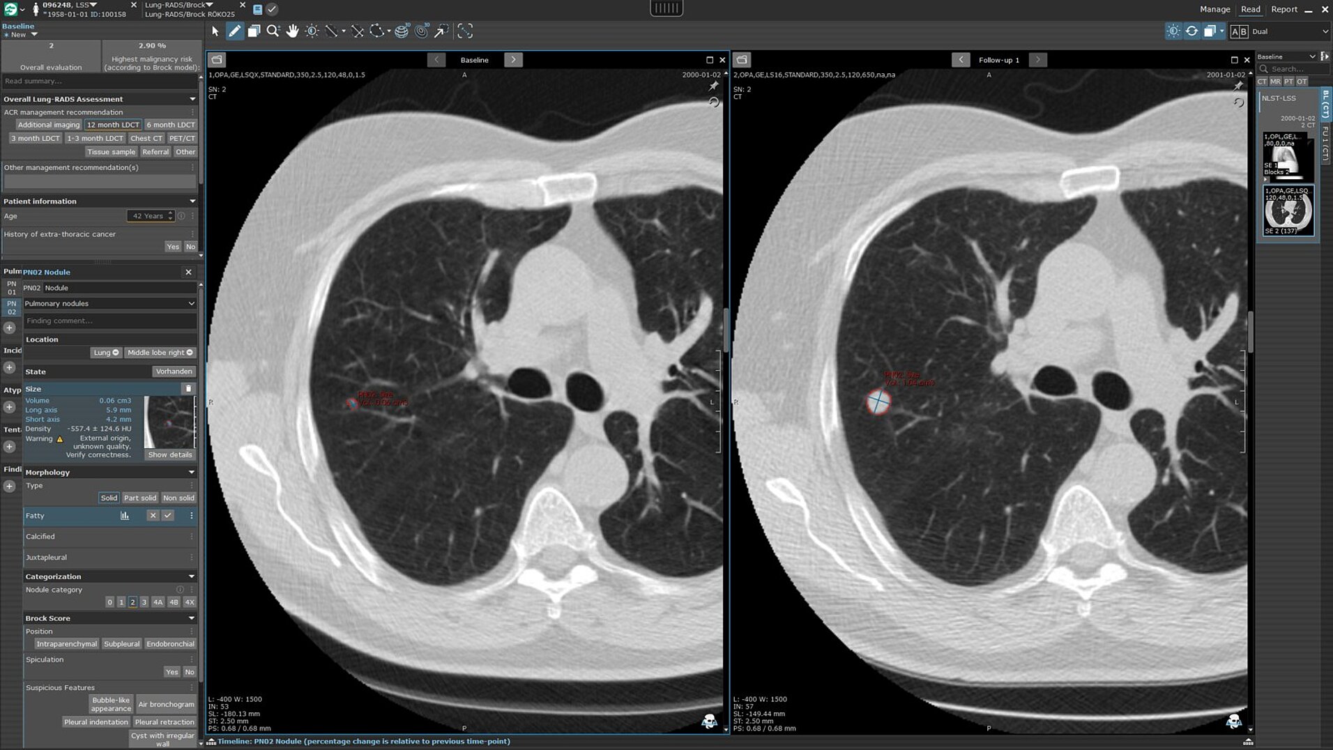

Guided reading
Built-in criteria and classification systems for screening, staging, and response evaluation, such as Lung-RADS, PI-RADS, TNM, RECIST 1.1, and RANO 2.0.

Longitudinal tracking
Images and prior measurements are synchronized, so each measured lesion is followed across examinations.

Automated structured reports
Reports with lesion snapshots, tables, and charts are generated directly from the read.

Radiomics
IBSI-compliant first- and second-order features from 2D and volumetric measurements, collected in the background.

Tumor growth rate modeling
Growth and regression rates modeled from longitudinal measurements give trial teams a complementary view of efficacy, potentially earlier and with fewer patients.

Data management and analytics
Exports in CSV, XML, PDF, and DICOM, FHIR-compliant data exchange, plus analytics dashboards.
From the first measurement to a reusable dataset: see mint Lesion in action with your own questions in mind.
How do radiologists and researchers use mint Lesion?
From daily reporting in the hospital to multi-center research projects, radiologists and researchers share how structured data in mint Lesion shapes their everyday practice. Discover more first-hand experiences in our success stories.
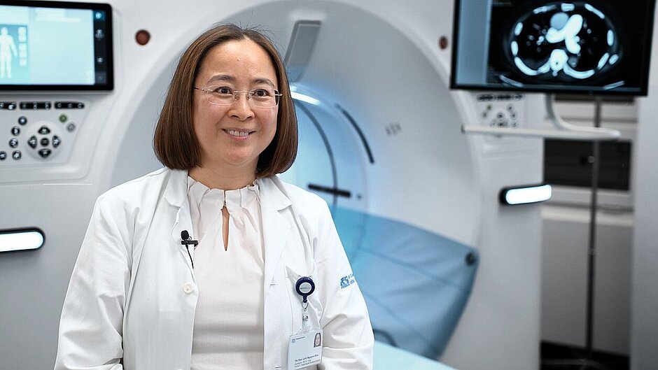
Structured Reporting in Clinical Practice
“For me, structured reporting in daily clinical practice is quality assurance.”
PD Dr. Thi Dan Linh Nguyen-Kim
Head of Department and Director, Institute of Radiology and Nuclear Medicine, Stadtspital Zürich
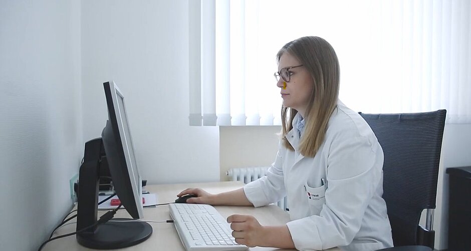
The Template Designer in Research Use
“In my view, this is its main advantage: the ability to collect clinical data in parallel with imaging data and directly relate the two.”
Dr. Madelaine Hettler
Resident physician, Sarcoma Center, University Medical Center Mannheim; RACOON-SAGA project (NUM)

Structured reporting and radiology workflows
Large multi-center study with imaging data and CRFs
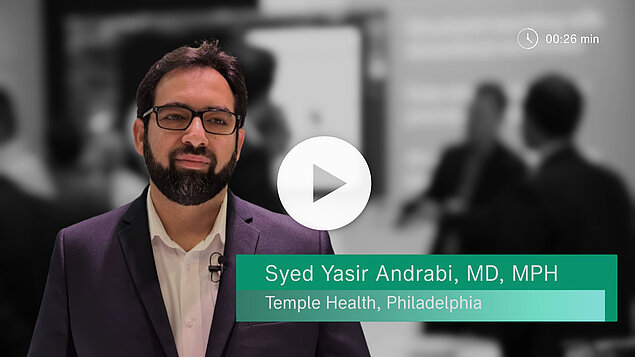
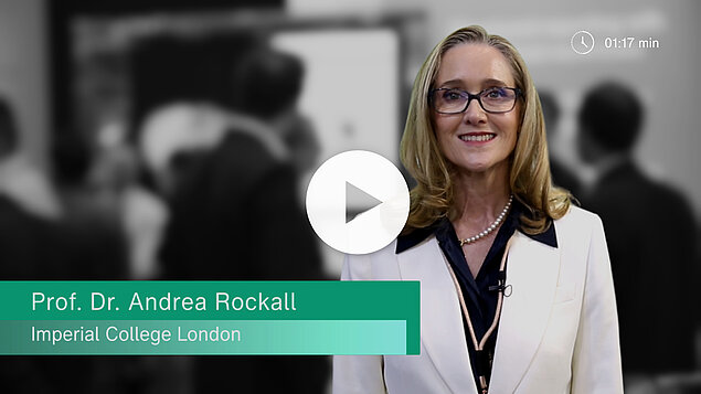
What does mint Lesion mean to you? One question, many perspectives: we invited mint Lesion users to share what the software means to them. Here are some of their answers.
How does mint Lesion make imaging data reusable beyond the read?
Every value recorded in mint Lesion is linked to its context: the examination, the finding it belongs to, the criteria applied, and the reader who documented it. Each read becomes structured, traceable data that can serve patient care, clinical trials, and research.

Captured in context
From the first measurement, each value is documented together with its origin.

Followed over time
Each measured lesion is followed across examinations, so data from every visit stays comparable.

Exported with integrity
Exports can carry raw data, intermediate data points, and user and approval information, digitally signed to support source data verification.

Reused beyond the read
Data captured in mint Lesion can be reused for further analysis and research. Radiomic features can be collected in the background.