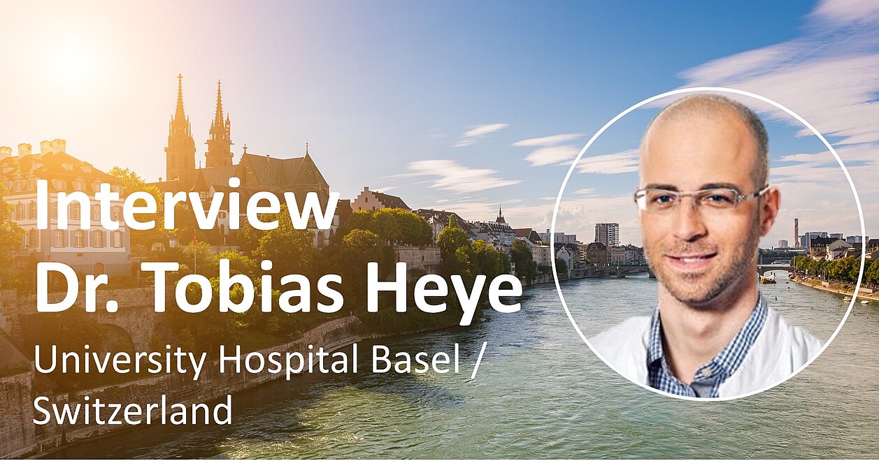
Since the end of 2011, mint Lesion™ has been in use at the University Hospital in Basel, Switzerland, – initially merely as a tool for clinical trials, but now also as the standard instrument for the entire oncologic radiology in clinical routine. We talked with PD Dr. Tobias Heye about his experiences with mint, the changes brought along with the use of the software, and his requests for future developments.
When you decided to use mint Lesion™ in Basel in 2011, what were the expectations you had for the product? Which problems and challenges did you want to address by using the software?
In 2011, I was not working at the University Hospital Basel yet, so I would like to refer to Prof. Bongartz here. The decision for mint Lesion™ was made for different reasons then. On the one hand, conducting tumor response assessments without structured reporting is very tedious and error-prone. The demonstration of radiologic results in an interdisciplinary context also requires a standardized documentation. This is where mint Lesion™ comes in, because unlike a normal radiologic PACS, the software offers exactly these possibilities of structured reporting and as a consequence, a clearly structured and comprehensible report. But Mint also acts in another direction, because a further challenge for radiologists is the fact that the set of rules for tumor follow-up assessments (RECIST, etc.) is very complex and requires a certain experience and attentiveness. In a training organization like ours, it is important to have these rules integrated directly into the reading interface in order to prevent mistakes and guarantee a consistency in the evaluation even in cases when the single time points are being assessed by different radiologists. All this is possible with mint Lesion™.
How is mint Lesion™ being used in Basel and how long did it take until it had established itself as a further reading tool?
In the beginning, mint was only used for single oncologic trials in the therapy response assessment, but for a bit more than two years now, mint has also been used for the reading of the entire oncologic image data. There are no exceptions to this, mint is the standard reading tool in oncology. We now use the software for second reads of external image data.
What is the main benefit of mint for you personally? What has changed the most in Basel thanks to the use of mint?
Structured reporting according to international oncologic standards is essential for the delivery of an adequate quality in the communication with referring oncologists. This is well supported in mint with the integrated reading profiles. At times where patients have up to three or four tumors in their patient history, a third or fourth line therapy, it is also important to keep an overview of the course of the disease. The graphic representation of the tumor development helps a lot in the comprehension and in the communication with the oncologists. The clear quality standards in oncologic reading procedures as well as the possibility of comprehensibly assessing the follow-ups are definitely the major changes brought along with the use of mint. mint has become a primary system in the radiologic oncologic reading procedure.
Where would you like to use mint Lesion™ beyond the current scope and which requirements arise from this?
For the future, we would like to have the possibility to continuously display the whole course from the initial staging to the follow-up assessment in one application, maintaining the history of tumor information (pre-/post-therapeutic staging etc.). Apart from that, the integration of clinical decision processes, e.g. decisions from tumor board reviews or laboratory parameters, would be necessary to use mint as a holistic oncologic platform. In our view, a function that allows the analysis of the underlying recorded data is necessary for the exploration of patterns in the tumor presentation and e.g. metastasis formation. It would also be desirable to have probabilities or a prediction based on the available data displayed in the software in order to be able to make a prognosis and estimate metastasis spread paths. Finally, the automatic segmentation and detection of lesions to support the assessment, including the displaying of the tumor burden, would be a huge leap in the development of the software.