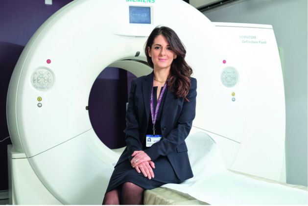
Since the beginning of 2017, all PI-RADS v2 reads are exclusively performed with mint Lesion™ by PD Dr. Matthias Röthke, Chief Medical Officer at Conradia in Hamburg. In this interview, he outlines the challenges of prostate cancer assessment and how mint supports this job-to-be-done.
What are the specific requirements for radiology when reading prostate images according to PI-RADS v2?
The PI-RADS system offers precise guidelines for the assessment of single lesions in the different sequences, i.e. T2 weighted imaging, diffusion weighted sequences and contrast enhanced perfusion. In addition to that, according to the PI-RADS system a distinction is made between lesions of the outlying peripheral prostate zone and nodal lesions of the transition zone located in the inner gland. Apart from the correct application of the scoring system, a special localization report of the findings needs to be given to the referring physician, usually a urologist, in the form of a standardized sector scheme.
How does mint Lesion™ help to fulfill these requirements?
Mint offers a continuous software assistance with the localization of possibly malignant lesions and their standardized documentation according to the PI-RADS scheme with the graphic embedment into the standardized prostate scheme. Furthermore, there are methods for volumetric measurements of the prostate. Another major advantage consists in the possibility to closely monitor patients in active surveillance, usually in case of a verified low-grade prostate carcinoma, longitudinally over the course of time and, if necessary, make an intervention or another biopsy of newly appeared/malignantly transformed lesions. Of course, the software is also perfectly suited for the assessment and documentation of patients in clinical trials.
How do urologists react to the new imaging result communication?
The urologists with whom I have been working with for several years explicitly welcome the standardized reporting and communication as well as the excellent result documentation using the sector scheme. The mint report has become the standard in the communication with the colleagues from urology who do not want to do without it anymore.
How can this development be evaluated from a patient’s perspective?
For patients, standardized reading procedures offer the greatest possible result certitude, because with the assistance of the software no important steps in the reading process of mpMRI or MR prostatography and in the determination of the respective PI-RADS scores can be overseen. In addition to that, the graphic result communication delivers precise information for the urologist to be able to biopsy a possibly suspicious lesion in a precise and targeted way. In this way, false negative biopsies can be avoided. For patients in active surveillance, the possibility of a standardized longitudinal follow-up assessment offers maximum accuracy and certitude.